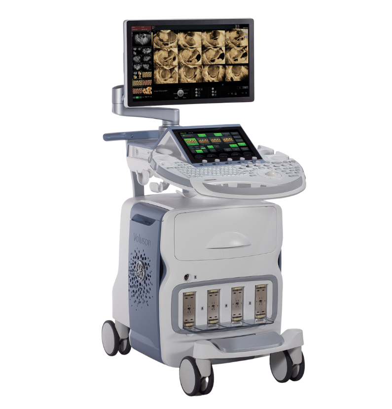
![Voluson™ E8 RSA Ultrasound System](/media/_processed_/5/4/csm_E8_small_banner_54bca6763f.png)
Voluson™ E8 RSA
Handle women’s health exams efficiently with ease and precision.
- Product Overview
- Imaging capabilities
- Downloads
Voluson E8 RSA
At a glance
Premium image quality
The powerful Radiance System Architecture provides faster processing speed and high frame rates, improving detail and contrast resolution.
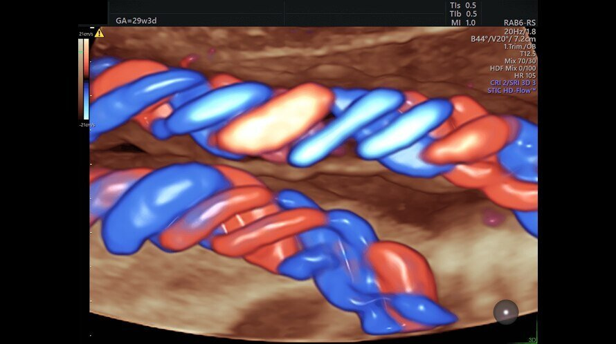
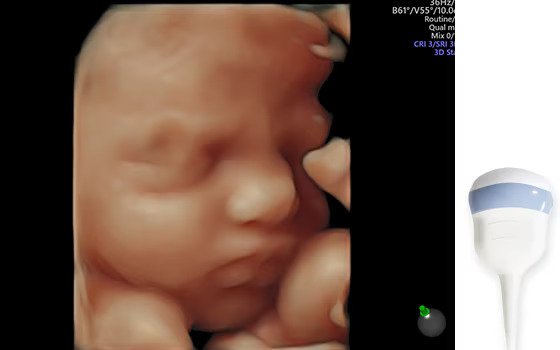
Comprehensive fetal heart tools
Benefit from advanced tools to assess and monitor the fetal heart. For example, with fetalHQ you can assess size, shape and contractility from the 4-chamber view.
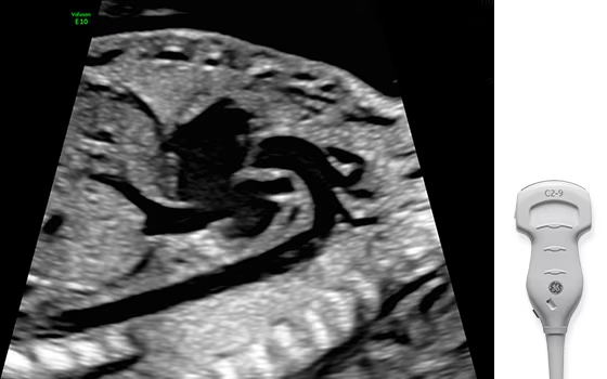
High performance XDclear™ probes
Achieve exceptional detail resolution with XDclear probe technology, offering extraordinary 2D imaging even in hard-to-scan patients.
AI-enhanced efficiency
Decrease exam complexity and streamline your workflow with easy-to-use automation tools including SonoCNS, an Edison™ AI application.
Voluson E8 RSA
Advanced features. Simplified workflow.
The Voluson E8 RSA ultrasound system is designed to keep pace with busy schedules, whether you are handling routine screening and examinations or focusing on your most complex cases. Achieve premium image quality and take advantage of Voluson E8 RSA's advanced capabilities to deliver exceptional care to your patients.
Product Highlights
Maximize efficiency with modern ergonomic design
![Front view of the Voluson E8 ultrasound system](/media/ultrasound/Products/Voluson/Voluson_E8_RSA/Webseite_Highlight__1_.png)
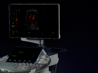
Excellent operator interface
Voluson E8 RSA is equipped with a 23" high-resolution monitor and a large 12.1" touch panel with Voluson xTouch functionality. The 1-button control panel allows for quick and convenient up and down.
AI-based automation features
A wide range of automation algorithms and other ultra-modern features enable flexible and efficient workflows.
Advanced beamformer design
Equipped with a high-quality beamformer, Voluson E8 RSA allows you to deliver ultrasound images with enhanced contrast resolution.
One touch responsiveness
The most commonly used scanning modes are available with a single keystroke.
Fast and secure data management
Use the Voluson E8's state-of-the-art communication technologies to quickly exchange data with patients and colleagues, and to archive your images easily. The integrated barcode scanner allows you to transfer patient information quickly and easily.
Product brochure
Discover detailed information about the Voluson E8 RSA ultrasound system and learn how it will help you to deliver confident care efficiently.
![](/whitepapers/_processed_/9/d/csm_300-19-U010E--Voluson-E8-BT19--sales-piece-_Aug_--JB59717XXh_37e63a252b.png)
For routine and complex exams
Advanced imaging technologies for diagnostic confidence
Voluson E8 RSA can help you stay at the forefront of innovation and deal with increasing patient volumes. The system's advanced beamformer design allows for enhanced contrast resolution, while the high-performance CPU guarantees fast processing speeds and frame rates. Thus you can confidently acquire the images you need, even in case of highly complex pathologies.
Voluson technology
Advanced ultrasound imaging with Voluson
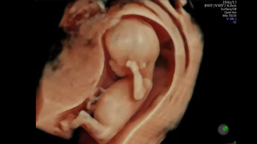
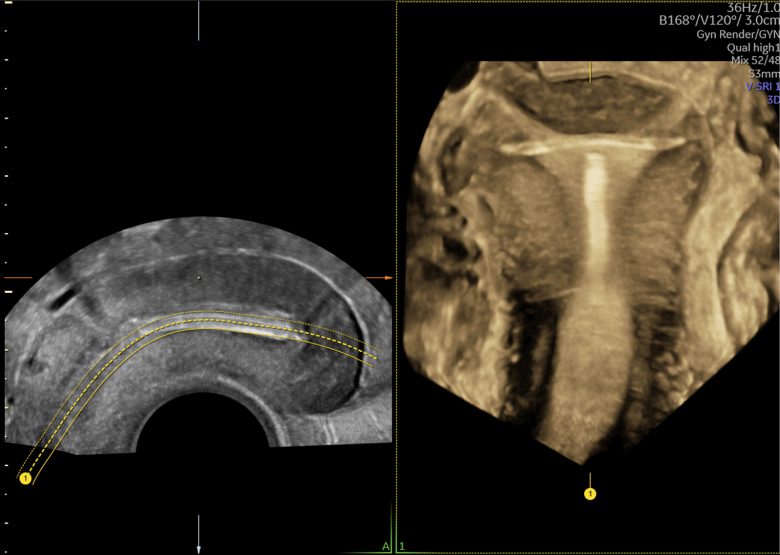
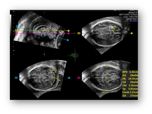
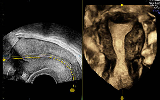
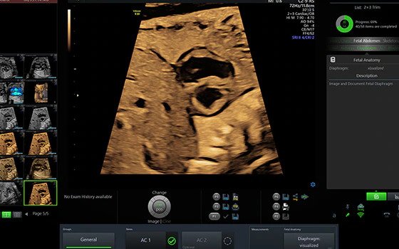
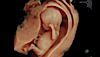
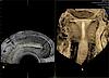
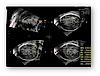
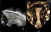
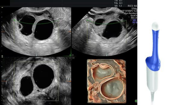
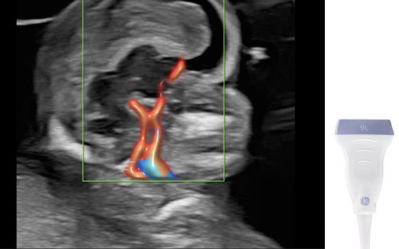
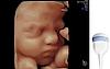
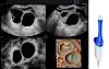
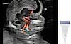
Radiantflow™: Achieve a new standard of color Doppler with Radiantflow - delivering easy, fast visualization even of the tiniest vessels.
Expand the range of visible blood flow to include low velocities with SlowflowHD to visualize blood perfusion.
![Ultrasound image of a fetus captured using HDlive technologies](/media/_processed_/1/4/csm_HDlive_Technologies_The_suite_of_HDlive%E2%84%A2_technologies_is_an_essential_problem_solving_tool_for_a_deeper_understanding_of_relational_anatomy_and_developing_structures._83352cdfbb.jpg)
The suite of HDlive™ technologies is an essential problem solving tool for a deeper understanding of relational anatomy and developing structures.
![Ultrasound image of an IUD captured using OmniView](/media/ultrasound/Products/Voluson/Voluson_Expert_22/omni-view-img.jpg)
Advanced VCI with OmniView: Streamline your volume imaging by obtaining any plane from a 3D or 4D volume by simply drawing a line, curve, poly-line, or trace. Further enhance image with adjustable slice thickness for improved contrast resolution.
![Ultrasound image of the fetal brain captured using SonoCNS](/media/ultrasound/Products/Voluson/Voluson_E8_RSA/SonoCNS.jpg)
SonoCNS is an Edison™ application using deep learning technology that helps you to properly align and display recommended views and measurements of the fetal brain.
![3D ultrasound image of the female uterus](/media/ultrasound/Products/Voluson/Voluson_E10/Uterine_Trace_Taking_the_effort_out_of_3D_with_Uterine_Trace%E2%80%94in_3_simple_steps__obtain_the_coronal_plane_of_the_uterus._Links_directly_to_Uterine_Classification_pictograms_for_easy_documentation_of_uteri.jpg)
Take the effort out of 3D with Uterine Trace, which acquires a volume of the uterus and extracts the coronal plane in a few simple steps. The direct link to Uterine Classification allows for easy documentation.
![Ultrasound image captured using Scan Assistant](/media/ultrasound/Products/Voluson/Voluson_E8_RSA/technology-scanassist.jpg)
Scan Assistant: This flexible, customizable exam protocol helps reduce time required to conduct and document results by guiding you through an exam more efficiently, aiding in annotation, measurement and reporting.
![](/_assets/b9df004e7f37d8e39e1f5da3327f12bc/Images/Generic/thumbnailposter.jpg)
![Ultrasound image of a fetus captured using HDlive technologies](/media/_processed_/1/4/csm_HDlive_Technologies_The_suite_of_HDlive%E2%84%A2_technologies_is_an_essential_problem_solving_tool_for_a_deeper_understanding_of_relational_anatomy_and_developing_structures._4ecc866261.jpg)
![Ultrasound image of an IUD captured using OmniView](/media/_processed_/f/7/csm_omni-view-img_7bb9099c91.jpg)
![Ultrasound image of the fetal brain captured using SonoCNS](/media/_processed_/7/8/csm_SonoCNS_2f8f35fa9f.jpg)
![3D ultrasound image of the female uterus](/media/_processed_/3/e/csm_Uterine_Trace_Taking_the_effort_out_of_3D_with_Uterine_Trace%E2%80%94in_3_simple_steps__obtain_the_coronal_plane_of_the_uterus._Links_directly_to_Uterine_Classification_pictograms_for_easy_documentation_of_uteri_08b1ecd58e.jpg)
![Ultrasound image captured using Scan Assistant](/media/_processed_/4/6/csm_technology-scanassist_595c637ba4.jpg)
Clinical images
Voluson E8 RSA at work
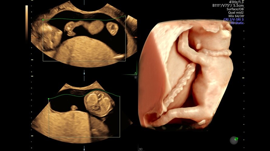
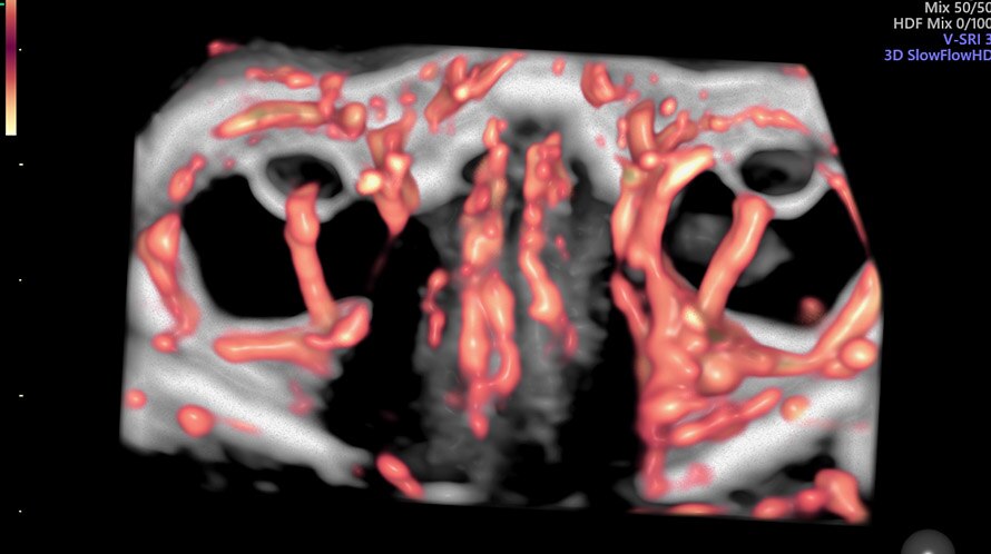
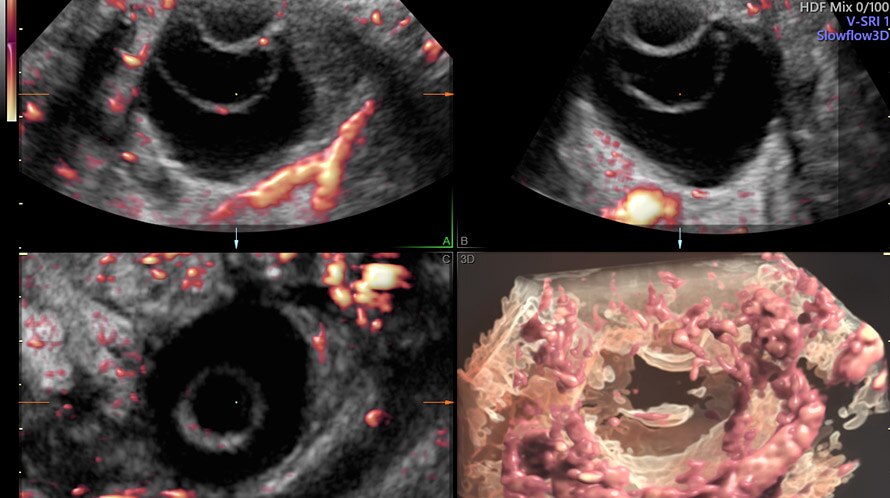
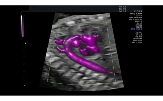
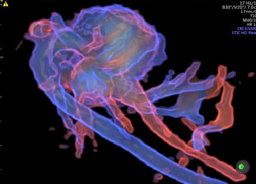
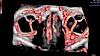
![Ultrasound image of a fetus captured using HDlive](/media/ultrasound/Products/Voluson/Voluson_E10/fetus-with-hdlive-generated-with-sonorenderlive.jpg)
Fetus with HDlive generated with SonoRenderlive.
![Ultrasound image of 19-week orbit captured using SlowFlow HD.](/media/ultrasound/Products/Voluson/Voluson_E10/19-week_orbit_with_Slowflow3D.jpg)
19-week orbit with Slowflow3D.
![Ultrasound image of an ovarian cyst captured using Slowflow3D](/media/ultrasound/Products/Voluson/Voluson_E10/gallery_ovarian-cyst-demonstrated-with-slowflow3d_pic3.jpg)
Ovarian cyst demonstrated with Slowflow3D
![Ultrasound image of the fetal heart captured using Radiantflow](/media/ultrasound/Products/Voluson/Voluson_E8_RSA/s10e-bt22---hdlive-umbilical-cord.jpg)
HDlive umbilical cord
![Ultrasound image of the aortic arch captured with Radiantflow](/media/ultrasound/Products/Voluson/Voluson_S8_Touch/radiantflow-aortic-arch.jpg)
Fetal aortic arch captured with Radiantflow
![Ultrasound image captured using HDlive Flow Silhouette](/media/ultrasound/Products/Voluson/Voluson_E8_RSA/HDlive-Flow-Silhouette-1.jpg)
HDlive Flow Silhouette
![Ultrasound image of a fetus captured using HDlive](/media/_processed_/a/e/csm_fetus-with-hdlive-generated-with-sonorenderlive_54d21d53a5.jpg)
![Ultrasound image of 19-week orbit captured using SlowFlow HD.](/media/_processed_/f/2/csm_19-week_orbit_with_Slowflow3D_bc485a7c26.jpg)
![Ultrasound image of an ovarian cyst captured using Slowflow3D](/media/_processed_/a/c/csm_gallery_ovarian-cyst-demonstrated-with-slowflow3d_pic3_1de571ec3f.jpg)
![Ultrasound image of the fetal heart captured using Radiantflow](/media/_processed_/b/e/csm_s10e-bt22---hdlive-umbilical-cord_cf6160242c.jpg)
![Ultrasound image of the aortic arch captured with Radiantflow](/media/_processed_/1/4/csm_radiantflow-aortic-arch_6716b7af16.jpg)
![Ultrasound image captured using HDlive Flow Silhouette](/media/_processed_/e/a/csm_HDlive-Flow-Silhouette-1_e49e64b5f2.jpg)
Clinical Images
Voluson probe technology
Exceptional image quality starts with advanced probe technology. Based on feedback from physicians and sonographers, Voluson probes are designed to meet your ergonomic requirements. Voluson probes use advanced technologies that enable you to deliver your best care for your patients.
![Ultrasound image captured with XDclear probe.](/media/ultrasound/Products/Voluson/Voluson_E8_RSA/probe-c2-9__2_.jpg)
Achieve exceptional tissue and detail resolution with XDclear probe technology C2-9-D and C1-6-D – the combination of single crystal, acoustic amplification and cool stack technology offers extraordinary 2D imaging even with difficult-to-scan patients.
![Ultrasound image captured using RAB6-D probe](/media/ultrasound/Products/Voluson/Voluson_E8_RSA/probe-rab6.jpg)
RAB6-D: An ultra-light volume probe with ergonomic grip and a thin lightweight cable for an easy acquisition of images.
![Ultrasound image captured using RIC5-9-RS probe](/media/ultrasound/Products/Voluson/Voluson_E8_RSA/probe-ric5.jpg)
RIC5-9-D: Wide band convex volume probe for first trimester and gynecological exams.
![Ultrasound image captured 9L High Frequency 2D](/media/ultrasound/Products/Voluson/Voluson_E8_RSA/9l_2dprobe.jpg)
9L is a wide-band high frequency 2D linear abdominal probe that helps you provide high quality 2D images.
![Ultrasound image captured with XDclear probe.](/media/_processed_/a/6/csm_probe-c2-9__2__dd240e74da.jpg)
![Ultrasound image captured using RAB6-D probe](/media/_processed_/2/a/csm_probe-rab6_5a4573f5ff.jpg)
![Ultrasound image captured using RIC5-9-RS probe](/media/_processed_/3/5/csm_probe-ric5_0f58c0a82b.jpg)
![Ultrasound image captured 9L High Frequency 2D](/media/_processed_/a/8/csm_9l_2dprobe_f52b104d88.jpg)
Dive into the world of Voluson E8 RSA
In this section you will find valuable and detailed information on Voluson E8 RSA and its features. Choose from brochures, whitepapers and case studies that offer you detailed insights into the functioning and application areas of our ultrasound system.
Product brochure
Discover detailed information about the Voluson E8 RSA ultrasound system and learn how it will help you to deliver confident care efficiently.
![](/whitepapers/_processed_/9/d/csm_300-19-U010E--Voluson-E8-BT19--sales-piece-_Aug_--JB59717XXh_37e63a252b.png)