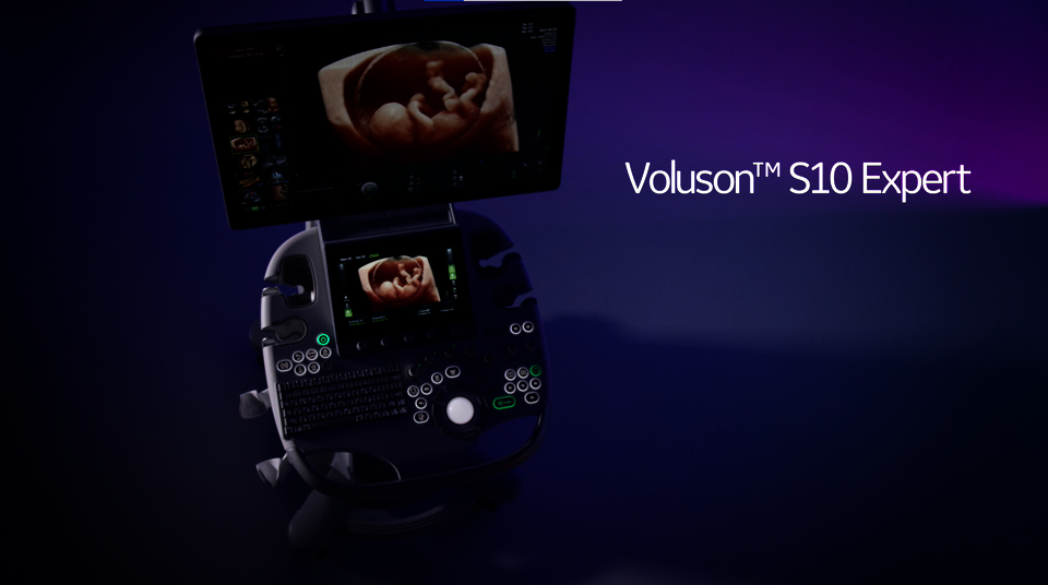
![The Voluson S10 Expert ultrasound system | GE HealthCare](/media/_processed_/5/2/csm_S10_expert_banner_small_745942761f.png)
Voluson™ S10 Expert
Powerful women’s health ultrasound system for your practice.
- Product Overview
- Imaging Capabilities
- Downloads
Voluson S10 Expert
At a glance
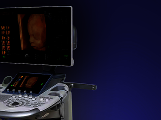
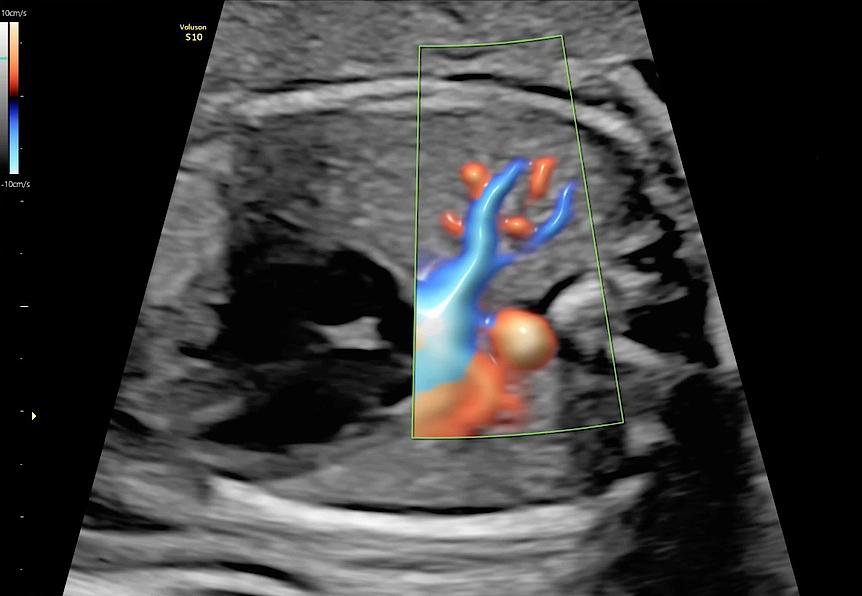
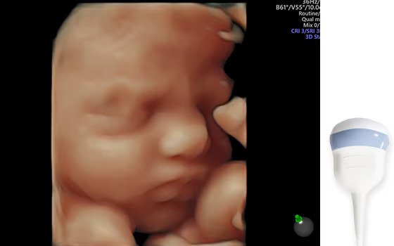
Advanced 3D/4D imaging
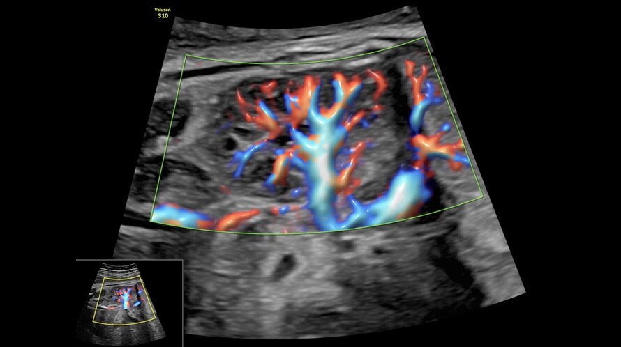
The powerful Voluson Core Architecture enables exceptional performance in all modes and drives innovative imaging technologies, including Radiantflow™ and XDclear™ probes.
Workflow Efficiency
Let Voluson’s automation technologies do the work for you. By automating tasks and calculations, you can save time, enhance consistency and improve accuracy.
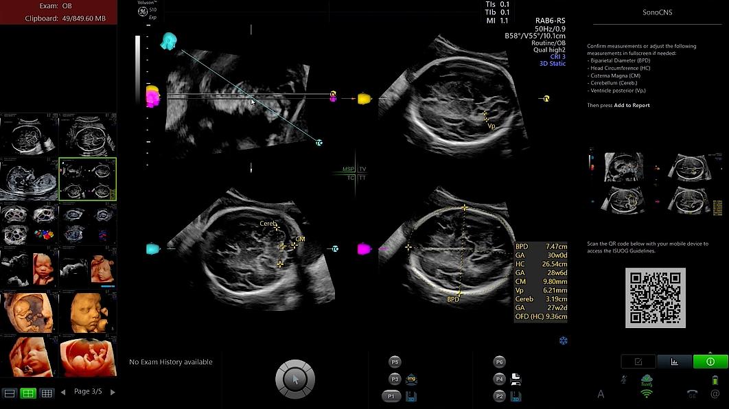
Expert capabilities made affordable
Experience industry-leading innovations migrated from our most advanced platform. Harness the power of AI-based tools like SonoCNS for accurate fetal brain diagnoses.
Comprehensive support
Leverage the entire Voluson ecosystem to access secure image archiving and reporting solutions, training resources and flexible service and support to keep your system secure and up-to-date.
Voluson S10 Expert
Get ahead. Stay ahead.
Take the next step forward with Voluson S10 Expert, the smart investment that inspires confidence with fast, clear, and consistent ultrasound imaging in 2D and 3D/4D. Scan with more ease and accelerate your workflow supported by GE HealthCare's industry-leading technology and advanced AI-based tools. Innovative features and additional resources from the Voluson ecosystem will help you to develop a reputation for excellence in women’s health – thus opening new pathways to clinical growth.
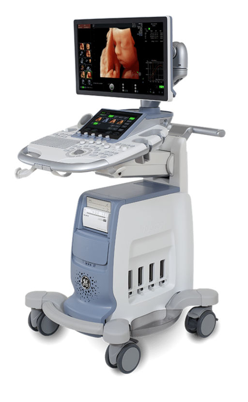
Product Highlights
Efficient workflows. Ergonomic design.
![Voluson S10 Expert ultrasound system](/media/ultrasound/Products/Voluson/Voluson_S10_Expert/S10_Expert__cropped_.png)
Fast and secure data management
Use the Voluson S10 Expert's state-of-the-art communication technologies to quickly exchange data with patients and colleagues and to archive your images easily.
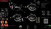
Ergonomic operator interface
Voluson S10 Expert is equipped with a large 10.1" touch panel with Voluson xTouch functionality for an intuitive volume navigation experience. The panel's horizontal and vertical position can be adjusted with just one touch of a button.
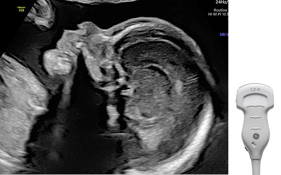
Advanced probe technology
Voluson S10 Expert comes with an ultra-light and high-frequency abdominal probe that is ideal for difficult examination conditions. In addition, a special transducer with matrix array technology is available for advanced mamma diagnostics.
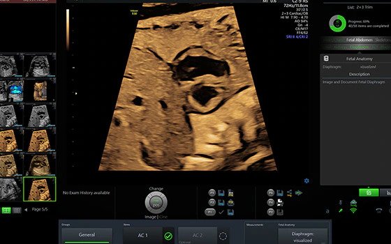
Intuitive and efficient design
Add TGC setting with a swipe and optimize it with just one keystroke. With Voluson S10 Expert, everything feels easy and familiar.
Four active probe ports
Voluson S10 Expert allows for quick and easy probe handling thanks to its four active probe ports.
Product brochure
Discover detailed information about the Voluson S10 Expert ultrasound system and learn how it will help you to deliver confident care efficiently.
![](/whitepapers/_processed_/2/7/csm_Product-sales-sheet--Voluson-S10E-BT22--JB18025XX-_2021__504affe714.png)
Expert ultrasound for your practice
Expert imaging capabilities made affordable
As your practice evolves, you need a dynamic system that grows with you. With the Voluson S10 Expert, you can take advantage of the industry-leading technology of our most advanced ultrasound platform at a more affordable price. Based on the Voluson Core Architecture, the S10 Expert will benefit from additional technological innovations as they become available.
Voluson technology
Discover Voluson technology for advanced ultrasound imaging
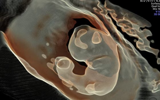
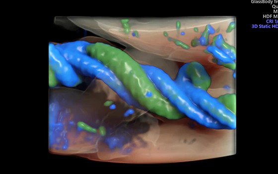
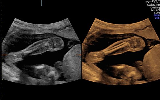
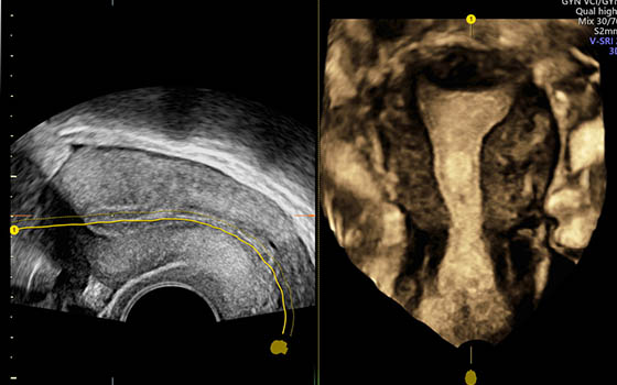
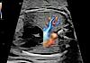
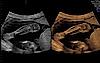
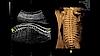
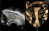
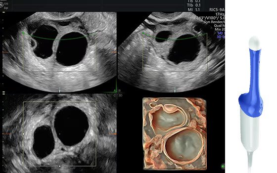
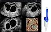
![Ultrasound image captured using Radiantflow](/media/_processed_/0/9/csm_radiantflow_77f2aee1a8.jpg)
Radiantflow™ demonstrates enhanced separation between vessels by adding height and depth information to provide a 3D-like appearance and the perception of dynamic blood flow.
![Ultrasound image of a fetus captured using HDlive Silhouette](/media/ultrasound/Products/Voluson/Voluson_S10_Expert/technology-hdlive-silhouette.jpg)
HDlive™ Silhouette: Dynamically apply targeted transparency to rendered structures for a more thorough view of anatomy from a solid surface structure to developing internal anatomy.
![Ultrasound image captured using HDlive Flow](/media/ultrasound/Products/Voluson/Voluson_S10_Expert/technology-hdlive-flow.jpg)
HDlive Flow displays vascular structures and orientation with greater dimension and illumination.
![Ultrasound image of the fetal brain captured using SonoCNS](/media/_processed_/9/a/csm_sonocns-11_11zon_b1f76848c4.jpg)
SonoCNS is an Edison™ application using deep learning technology that helps you to properly align and display recommended views and measurements of the fetal brain.
![Ultrasound image captured using Volume Contrast Imaging (VCI)](/media/ultrasound/Products/Voluson/Voluson_S10_Expert/technology-vci.jpg)
Volume Contrast Imaging (VCI) adjusts slice thickness on 3D or 4D images by using bone and tissue rendering techniques to help enhance contrast resolution.
![Ultrasound image of the fetal spine](/media/ultrasound/Products/Voluson/Voluson_S8_Touch/OmiView.jpg)
OmniView: Obtain any view from a volume by drawing a line, curve, poly-line, or trace through a structure enabling views of irregularly shaped structures.
![3D ultrasound image of the female uterus](/media/ultrasound/Products/Voluson/Voluson_E10/Uterine_Trace_Taking_the_effort_out_of_3D_with_Uterine_Trace%E2%80%94in_3_simple_steps__obtain_the_coronal_plane_of_the_uterus._Links_directly_to_Uterine_Classification_pictograms_for_easy_documentation_of_uteri.jpg)
Take the effort out of 3D with Uterine Trace, which acquires a volume of the uterus and extracts the coronal plane in a few simple steps. The direct link to Uterine Classification allows for easy documentation.
![Ultrasound image captured using Scan Assistant](/media/ultrasound/Products/Voluson/Voluson_S10_Expert/technology-scanassist.jpg)
Scan Assistant: This flexible, customizable exam protocol helps to reduce time required to conduct and document results by guiding you through an exam more efficiently and by aiding in annotation, measurement and reporting.
![Ultrasound image captured using Radiantflow](/media/_processed_/0/9/csm_radiantflow_bcffa99a3a.jpg)
![Ultrasound image of a fetus captured using HDlive Silhouette](/media/_processed_/d/b/csm_technology-hdlive-silhouette_db6fbb5a2d.jpg)
![Ultrasound image captured using HDlive Flow](/media/_processed_/a/8/csm_technology-hdlive-flow_d2e22097ca.jpg)
![Ultrasound image of the fetal brain captured using SonoCNS](/media/_processed_/9/a/csm_sonocns-11_11zon_f8d3ef8847.jpg)
![Ultrasound image captured using Volume Contrast Imaging (VCI)](/media/_processed_/3/8/csm_technology-vci_f11cedc80e.jpg)
![Ultrasound image of the fetal spine](/media/_processed_/a/c/csm_OmiView_dd6932a219.jpg)
![3D ultrasound image of the female uterus](/media/_processed_/3/e/csm_Uterine_Trace_Taking_the_effort_out_of_3D_with_Uterine_Trace%E2%80%94in_3_simple_steps__obtain_the_coronal_plane_of_the_uterus._Links_directly_to_Uterine_Classification_pictograms_for_easy_documentation_of_uteri_08b1ecd58e.jpg)
![Ultrasound image captured using Scan Assistant](/media/_processed_/6/2/csm_technology-scanassist_223ca374d2.jpg)
Clinical images
Voluson S10 Expert at work
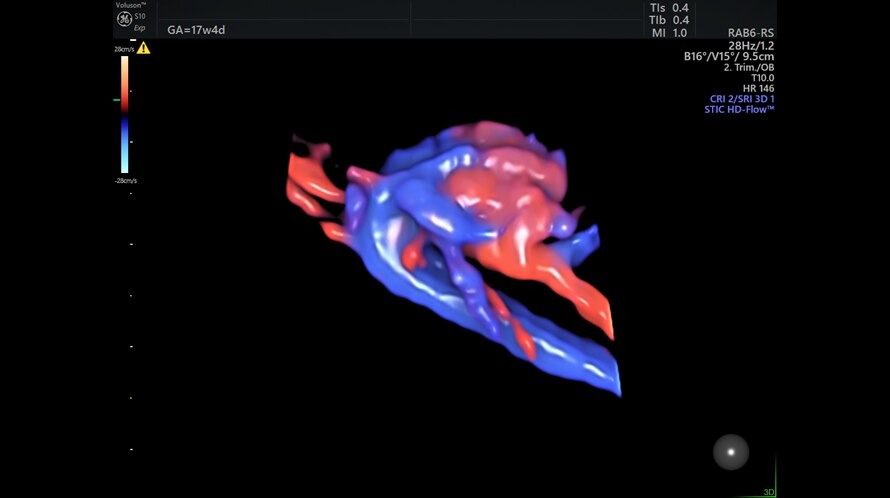
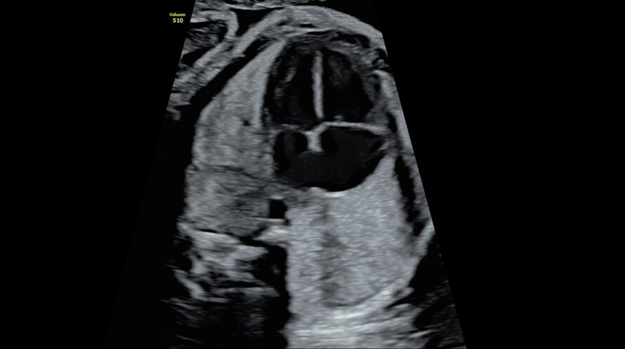
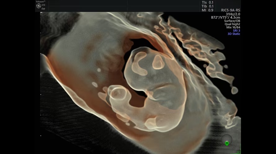
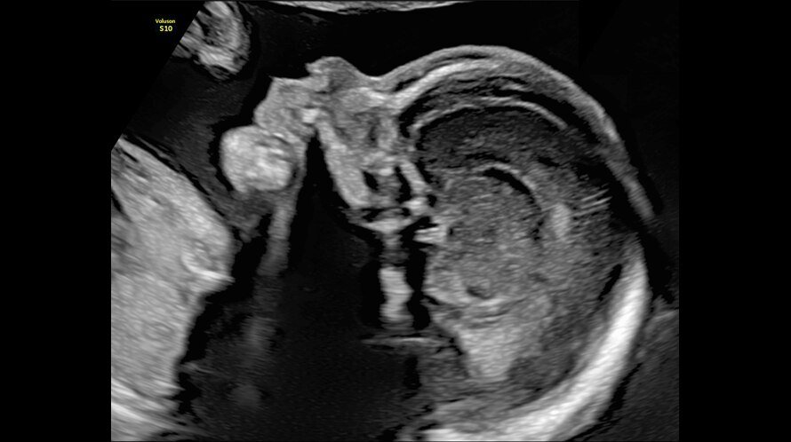
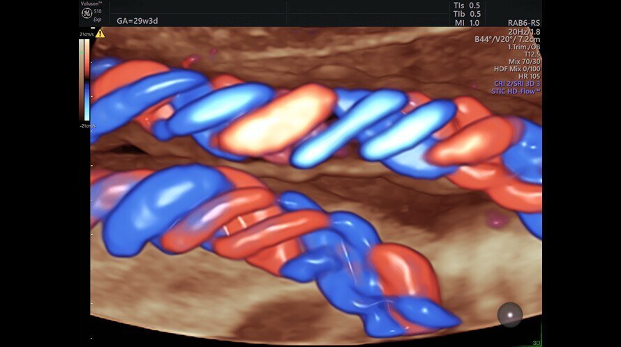
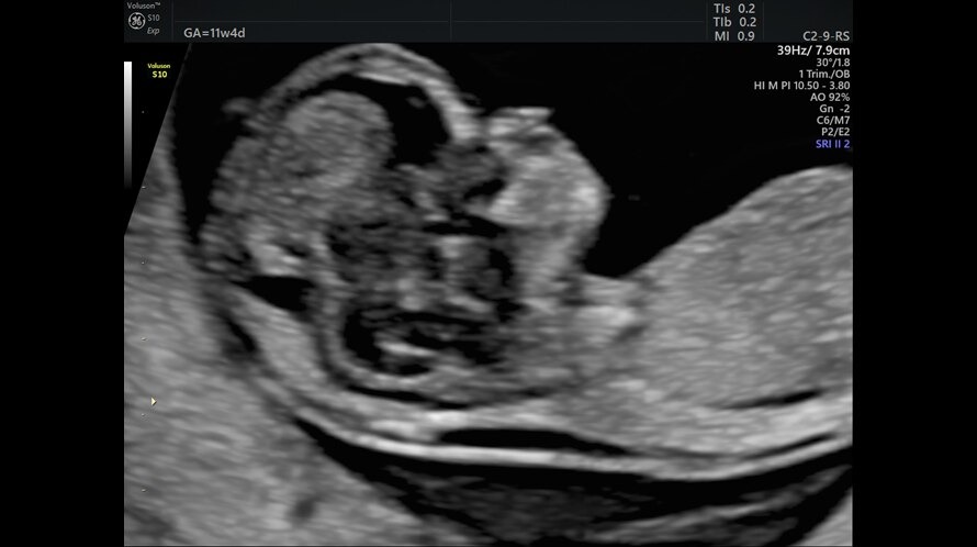
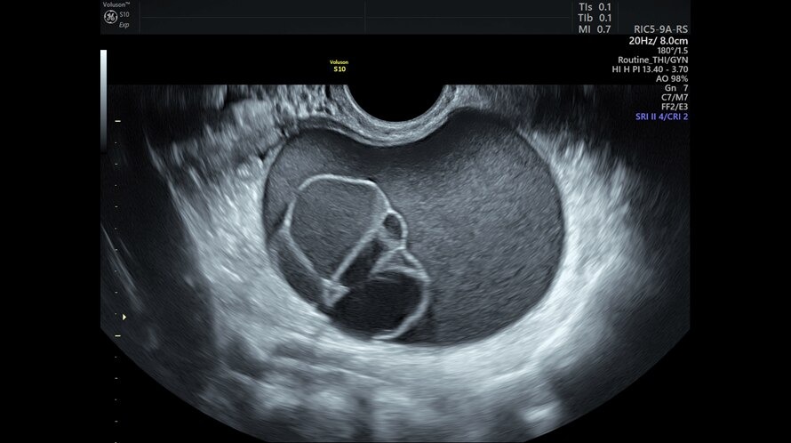
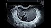
![Ultrasound image captured using HD Flow](/media/ultrasound/Products/Voluson/Voluson_S10_Expert/stic_with_hd-flow_fetal_11zon.jpg)
STIC with HD-Flow™ fetal heart & aortic arch
![Ultrasound image of a fetal 4-chamber heart](/media/ultrasound/Products/Voluson/Voluson_S10_Expert/4-chamber_heart_11zon.jpg)
4-chamber heart
![Ultrasound image of an 8 week fetus captured using HDlive Silhouette](/media/ultrasound/Products/Voluson/Voluson_S10_Expert/hdlive_8_week_fetus_11zon.jpg)
HDlive Silhouette 8-week fetus
![Ultrasound image of a fetal profile](/media/ultrasound/Products/Voluson/Voluson_S10_Expert/fetal_profile.jpg)
Fetal profile
![Ultrasound image of the fetal heart captured using Radiantflow](/media/ultrasound/Products/Voluson/Voluson_S10_Expert/s10e-bt22---hdlive-umbilical-cord.jpg)
HDlive Flow umbilical cord
![Ultrasound image of a fetus](/media/ultrasound/Products/Voluson/Voluson_S10_Expert/nuchal_transulency_11zon.jpg)
Nuchal translucency
![Ultrasound image complex ovarian mass](/media/ultrasound/Products/Voluson/Voluson_S10_Expert/uterus_11zon.jpg)
Complex ovarian mass
![Ultrasound image of a fetal kidney captured using Radiantflow](/media/ultrasound/Products/Voluson/Voluson_S10_Expert/radiant_flow_fetal_kidney.jpg)
Radiantflow fetal kidney
![Ultrasound image captured using HD Flow](/media/_processed_/4/4/csm_stic_with_hd-flow_fetal_11zon_0e33725fef.jpg)
![Ultrasound image of a fetal 4-chamber heart](/media/_processed_/7/4/csm_4-chamber_heart_11zon_addae701f8.jpg)
![Ultrasound image of an 8 week fetus captured using HDlive Silhouette](/media/_processed_/b/b/csm_hdlive_8_week_fetus_11zon_4f549e63ec.jpg)
![Ultrasound image of a fetal profile](/media/_processed_/7/3/csm_fetal_profile_313036aee6.jpg)
![Ultrasound image of the fetal heart captured using Radiantflow](/media/_processed_/2/7/csm_s10e-bt22---hdlive-umbilical-cord_b2ee2fc1fa.jpg)
![Ultrasound image of a fetus](/media/_processed_/7/5/csm_nuchal_transulency_11zon_ffb73facdc.jpg)
![Ultrasound image complex ovarian mass](/media/_processed_/3/5/csm_uterus_11zon_5b0bab0522.jpg)
![Ultrasound image of a fetal kidney captured using Radiantflow](/media/_processed_/3/6/csm_radiant_flow_fetal_kidney_bfc0d270bc.jpg)
Clinical images
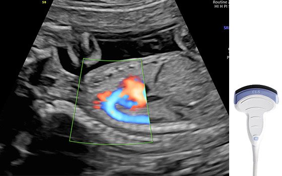
Voluson probe technology for extraordinary ultrasound images
Extraordinary image quality begins with advanced probe technology. Based on feedback from physicians and sonographers, Voluson probes have evolved to help meet your ergonomic needs and include innovations and advanced technology for women’s healthcare applications.
![Ultrasound image of fetal profile captured with C2-9-RS probe](/media/ultrasound/Products/Voluson/Voluson_S10_Expert/probe-c2-9.jpg)
C2-9-RS: Achieve exceptional tissue and detail resolution with C2-9 single crystal technology XDclear™ probe.
![Ultrasound image captured with C1-5-RS probe](/media/ultrasound/Products/Voluson/Voluson_S10_Expert/probe-c1-5rs.jpg)
C1-5-RS: Delivers excellent image quality and penetration when scanning larger patients.
![Ultrasound image captured using RAB6-RS probe](/media/ultrasound/Products/Voluson/Voluson_S10_Expert/probe-rab6.jpg)
RAB6-RS: An ultra-light volume probe with ergonomic grip and a thin lightweight cable for an easy acquisition of images.
![Ultrasound image captured using RIC5-9-RS probe](/media/ultrasound/Products/Voluson/Voluson_S10_Expert/probe-ric5.jpg)
RIC5-9-RS: Wide band convex volume probe for first trimester and gynecological exams.
![Ultrasound image of fetal profile captured with C2-9-RS probe](/media/_processed_/6/2/csm_probe-c2-9_386d136e77.jpg)
![Ultrasound image captured with C1-5-RS probe](/media/_processed_/5/a/csm_probe-c1-5rs_0bf88f5759.jpg)
![Ultrasound image captured using RAB6-RS probe](/media/_processed_/f/a/csm_probe-rab6_99e1da96c8.jpg)
![Ultrasound image captured using RIC5-9-RS probe](/media/_processed_/0/2/csm_probe-ric5_ec1b95b514.jpg)
Dive into the world of Voluson S10 Expert
In this section you will find valuable and detailed information on Voluson S10 Expert and its features. Choose from brochures, whitepapers and case studies that offer you detailed insights into the functioning and application areas of our ultrasound system.
Product brochure
Discover detailed information about the Voluson S10 Expert ultrasound system and learn how it will help you to deliver confident care efficiently.
![](/whitepapers/_processed_/2/7/csm_Product-sales-sheet--Voluson-S10E-BT22--JB18025XX-_2021__504affe714.png)