At a glance
Increased sensitivity
Use Invenia ABUS 2.0 as a complement to mammography and increase sensitivity in detecting breast cancer in dense breast tissue by 57%¹.
Advanced 3D imaging
Benefit from a complete 3D view of the breast, including the coronary plane to detect architectural distortions and multifocal disease rapidly.
Fast and efficient workflow
With Invenia ABUS 2.0, a complete review can be conducted in only 3 minutes² – an enormous saving of time compared to traditional breast ultrasound.
Optimal reproducibility
Thanks to standardized workflows and intelligent imaging algorithms, Invenia ABUS 2.0 offers exceptional image quality and cross-operator reproducibility.
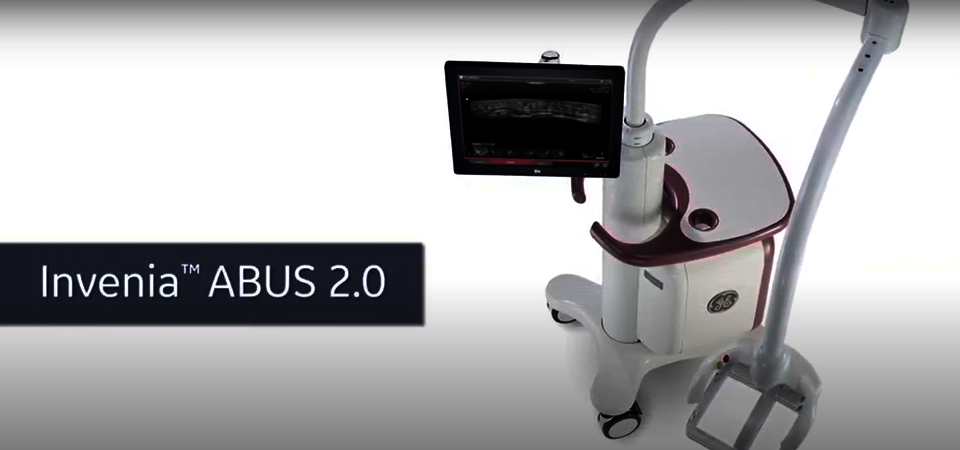
Invenia ABUS 2.0
Improve breast cancer detection
About 40% of all women around the world have dense breast tissue³. From a medical point of view, this is associated with a four to six times higher risk of breast cancer⁴. At the same time, traditional mammography is stretched to its limits when dealing with dense breasts - about 33% of cancers in dense breasts are missed⁵.
That's exactly why GE HealthCare has invented the Invenia ABUS 2.0, an ultrasound system specifically designed for the detection of cancer in dense breast tissue. Used as a supplement to mammography, Invenia ABUS 2.0 uses non-ionizing radiation and offers automated volumetric 3D ultrasound scanning of the breast, including image optimisation for accurate and reproducible results. The diagnosis of the acquired images usually takes no more than a few minutes.
Invenia ABUS 2.0
See dense breast tissue with clarity
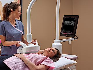
Invenia ABUS 2.0. in your daily workflow
Having received a brief training by our ultrasound experts, the examination with Invenia ABUS 2.0 can be carried out by your medical staff. Within an average scanning time of 15 minutes, the patient receives a comprehensive image of her breast architecture. Afterwards, the generated images are sent to the ABUS Viewer and reviewed by a physician. Usually, a complete review takes no more than 3 minutes.
Thanks to ABUS's advanced technology, 3D volumes are represented in the patented 2 mm coronary layer from the skin to the chest wall. The result: a diagnostic option that enables a quick and intuitive analysis even with difficult breast anatomy and pathology.
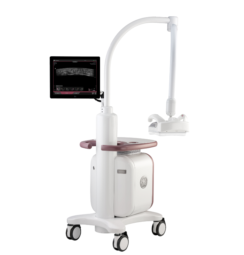
Product Highlights
Proactive and personalized breast care
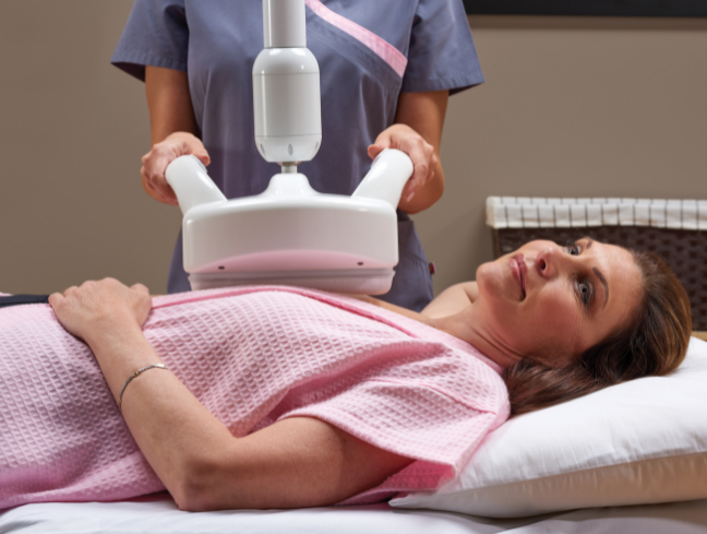
Designed for patient comfort
The high-frequency Reverse Curve™ transducer uses non-ionizing radiation and is shaped to match a woman’s anatomy, thus enabling complete coverage of the breast tissue. The consistent and homogeneous compression increases the examination comfort for the patient.
User-friendly interface
Equipped with a high resolution touchscreen and an ergonomic, graphical user interface, Invenia ABUS 2.0 guides you through the examination, thus ensuring a simple and smooth workflow.
Image optimization at the touch of a button
Use intelligent imaging algorithms to minimize artifacts at the touch of a button to achieve exceptional image quality and cross-operator reproducibility.
Powerful imaging architecture
Invenia ABUS 2.0 uses cSound™ Imageformer technology to create meaningful volumes with excellent image quality. The conventional parameters of hand-held ultrasound, such as focus zones and gain, are automatically optimised. Each pixel is therefore automatically in focus.
Efficient diagnosis via ABUS Viewer
The generated images are sent to the ABUS Viewer, which is designed for a fast and effective workflow. 3D volumes are represented in the patented 2 mm coronary layer from the skin to the chest wall. The result: A diagnostic option that enables a quick and intuitive analysis even with difficult breast anatomy and pathology.
Education and implementation support
To ensure smooth and seamless integration of Invenia ABUS 2.0 in your clinical workflow, GE HealthCare has developed a comprehensive implementation concept.
Training for physicians
Our Mastery Program for physicians is led by experienced and certified peer educators and helps quickly gain confidence in reading Invenia ABUS 2.0 images.
Training for medical professionals and technicians
During the installation, an ABUS Application Specialist will conduct a three-day training on-site on ultrasound technology, anatomy, ABUS scanning and quality analysis.
Marketing support
Real-world digital marketing tools and professionally designed templates are available. Recommendations for educating key audiences, workflow options and marketing strategies help you launch your ABUS program.


References
- Wilczek, Leifland, et. al. Adding 3D Automated Breast Ultrasound to mammography screening in women with heterogeneously and extremely dense breasts. European Journal of Radiology 85 (2016) 1554–1563
- Blumen, et. al. Comparison of treatment costs for breast cancer by tumor stage, and type of service.
- Pisano, E.D., Gatsonis, C., et. al. Diagnostic Performance of Digital versus Film Mammography for Breast-Cancer Screening, N Engl J Med 353 (2005) 1- 11
- Boyd, N.F., Guo, H., et. al., Mammographic Density and the Risk and Detection of Breast Cancer N Engl J Med 356 (3; 2007) 227-2362
- Kolb, T., Lichy, J., Newhouse, J.H., Comparison of the Performance of Screening Mammography, Physical Examination, and Breast US and Evaluation of Factors that Influence Them: An Analysis of 27,825 Patient Evaluations Radiology 225 (2002) 165–175
For better breast care
Improve early detection, treatment and monitoring
Invenia ABUS 2.0 supports you and your patients along the whole breast care pathway – from screening to diagnosis and staging to monitoring. As a first step, the ultrasound system is a valuable addition to your screening capabilities, increasing sensitivity in detecting breast cancer in dense breast tissue by 57%¹ when used in addition to mammography.
Once diagnosed, ABUS allows for more precise treatment planning as the multiplanar views provide a complete 3D breast image with reproducible localisation of pathological findings. Finally, Invenia ABUS 2.0 allows you to closely monitor the success of neoadjuvant chemotherapy.
All this is made possible by the ultrasound systems' intelligent imaging algorithms, including Tissue Equalisation, Nipple Shadow Compensation, Breast Border Detection and Chest Wall Detection. These algorithms serve to minimize artefacts and increase cross-operator reproducibility.
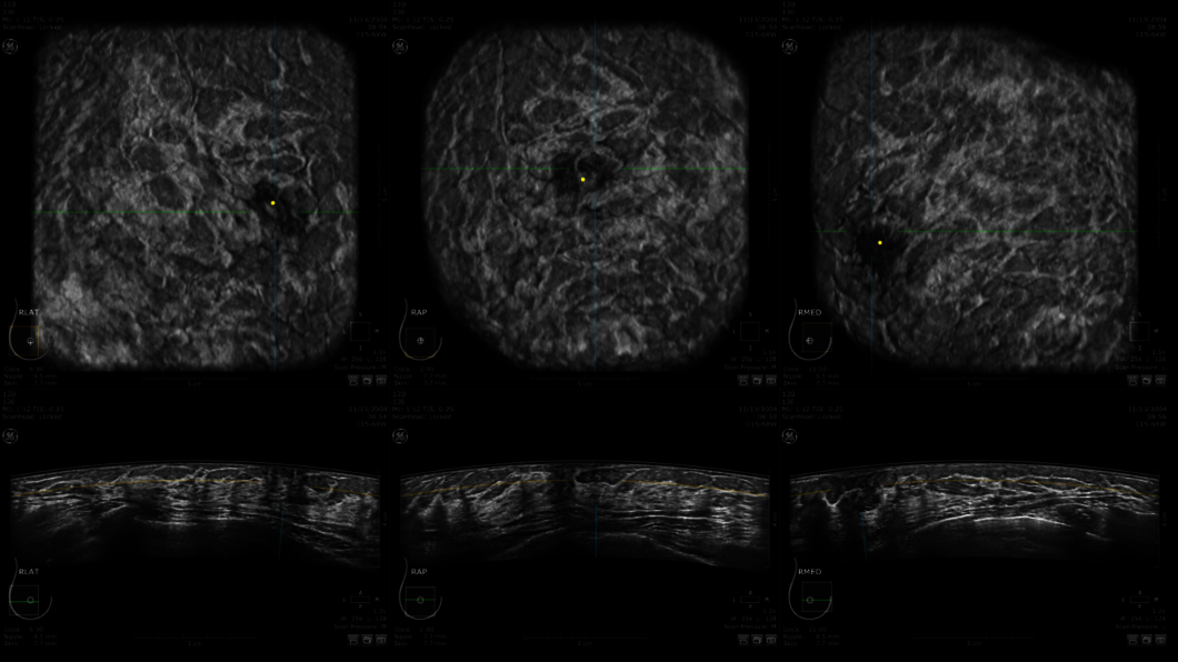
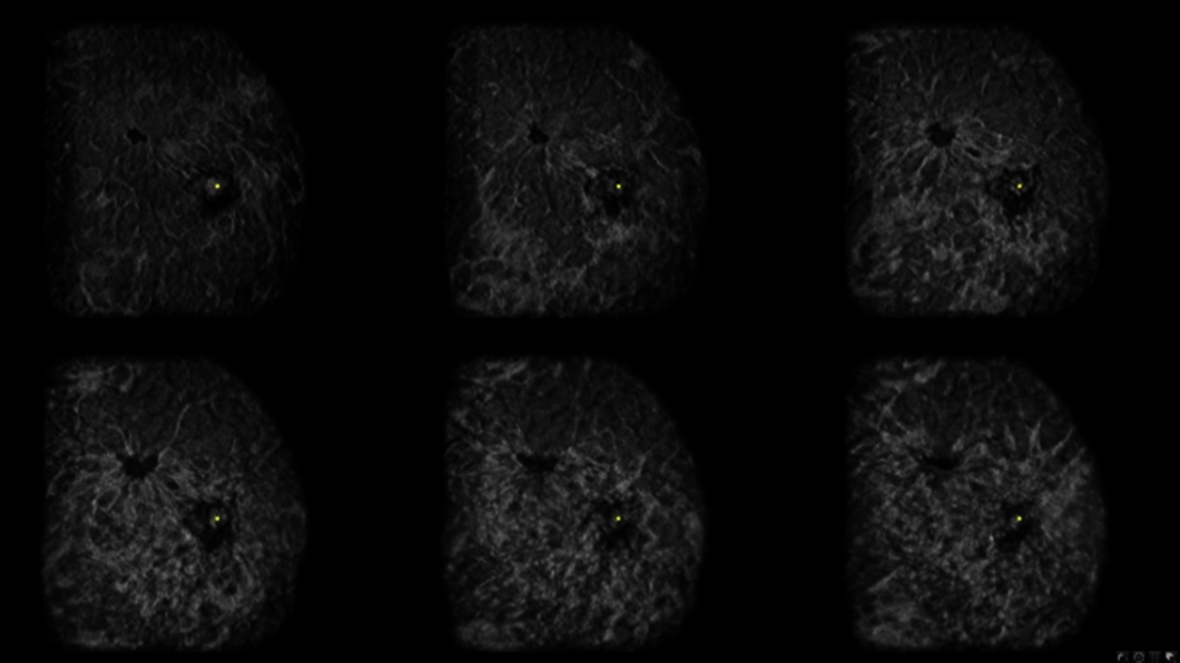
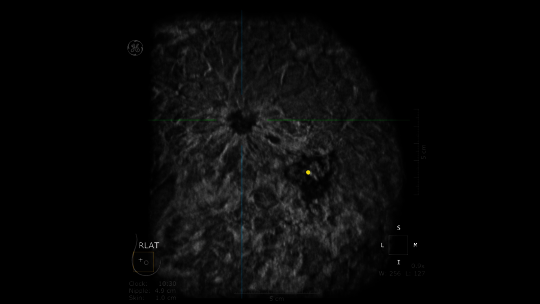
Clinical Images
Invenia ABUS 2.0 at work


References
- Wilczek, Leifland, et. al. Adding 3D Automated Breast Ultrasound to mammography screening in women with heterogeneously and extremely dense breasts. European Journal of Radiology 85 (2016) 1554–1563
Dive into the world of Invenia ABUS 2.0
In this section you will find valuable and detailed information on Invenia ABUS 2.0 and its features. Choose from brochures, whitepapers and case studies that offer you detailed insights into the functioning and application areas of our ultrasound system.


Painless. Quick. Peace of mind.
Agla, a nurse working in Affidea in Lithuania about patients' experience with Invenia ABUS 2.0

Many patients imagine breast scanning is painful, disturbing and time consuming.
Grazina, patient from Affidea in Vilnius about her experience with Invenia ABUS 2.0

Improve breast cancer outcomes through innovative imaging technology.
Dr Rūta Briedienė explains how Invenia ABUS 2.0 is changing Breast Care in Affidea clinic in Vilnius
Transforming breast care from passive to active.
Empower females to take control of their breast health and make more impact now.