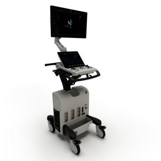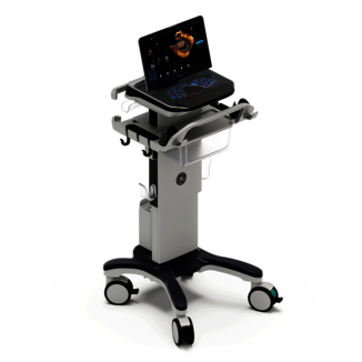
Interventional Cardiology
Discover superb visualization of the anatomy and seamless workflow for your
interventional procedures with Vivid™ Ultra Edition.
Demand for minimally invasive procedures is growing.
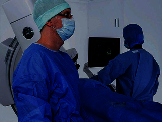
Structural heart procedure success depends on preparation, collaboration and clear communication within the heart team.
Your complex interventions may often require the use of fluoroscopy, CT and echo images to plan, guide and assess procedures, which may generate room for uncertainty & lack of clarity, potentially reducing efficiency and increasing risk for suboptimal interventions.
We are addressing all your needs
Optimized integration
Complex interventions require the use of several modalities in addition to ultrasound for planning and guidance. A well-thought-out ergonomics for these enviroments is paramount in order to reduce manipulations to the most effective actions, for optimized and safer procedures.
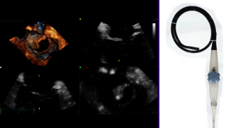
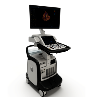
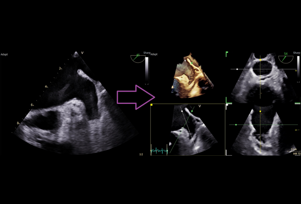
Dedicated Probes
A key element for an exceptional image quality is cSound™ software beamformer. The world's most compact mini-4D probe is suitable for a wide range of cardiology procedures.
Powerful Modeling
Simplification of live guidance and improved quality of communication within the cardiac team with CT Fusion.
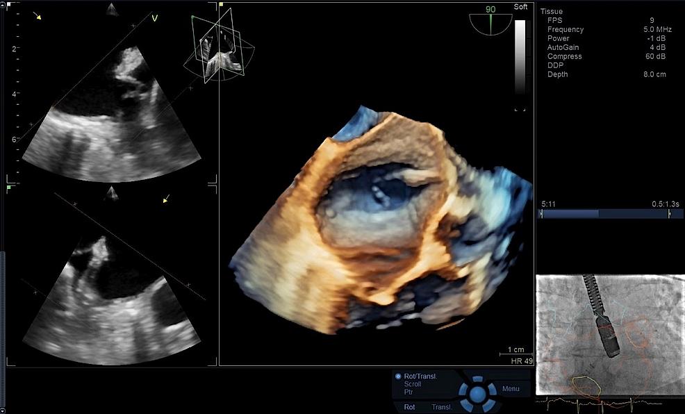
Precise visualization & guidance
Photorealistic visualization of anatomy with FlexiLight and HD color flow rendering technique for a simplified live guidance and improved quality of communication within the cardiac team with CT Fusion, 4D markers, FlexiSlice, and View-X.
Clinical Images
Exploring the impact of interventional Ultrasound Imaging in modern medicine

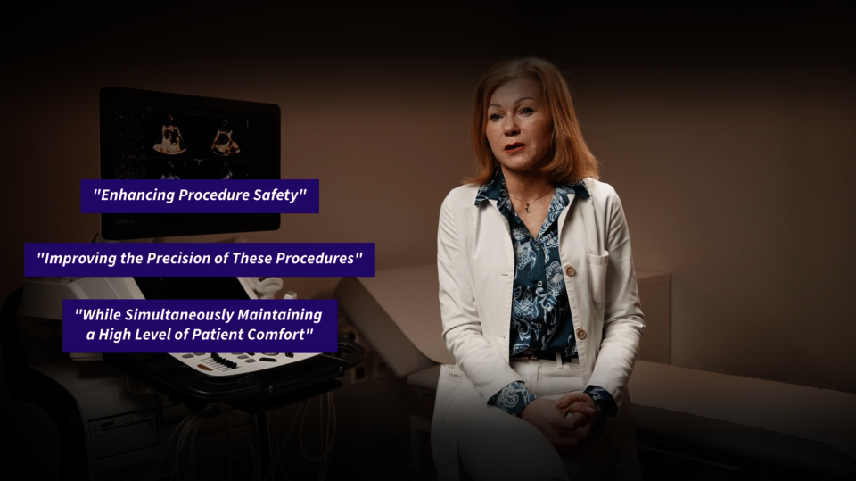
Clinical Expert’s perspective - Prof. Marcin Fijałkowski, MD, PhD
Patient comfort and safety with the 9VT-D mini 3D TEE Probe
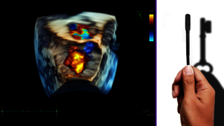
AI Flexiviews LAA
With a single click, AI Flexiviews LAA delivers AI-driven 4D visualization of the LAA. Trained to detect the LAA in 2D ultrasound data, its advanced algorithm instantly generates a 4D enface view and provides immediate access to FlexiSlice views for LAA assessment with minimal manual adjustments.
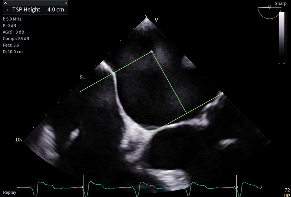
TSP Height
TSP Height tool helps you quickly find the ideal puncture location according to the guideline of the ongoing interventional procedure.
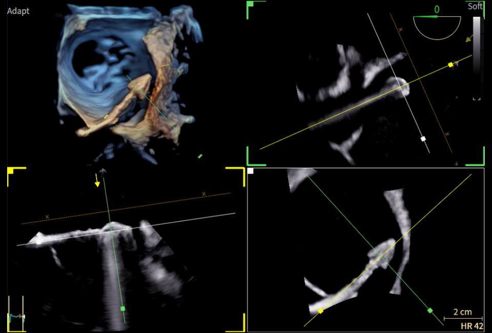
2-Click Align
Quick and easy alignment of FlexiSlice views along 3D structures, such as catheters, regurgitant jets or valves.
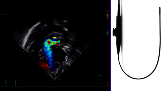
FlexiLight
Rendering techniques for photo-realistic
light-source based illumination of heart
structures, providing comprehensive
visualization of cardiac structures.
FlexiLight may allow a comprehensive
visualization of leaflets and regurgitant
orifices while ensuring the proper
alignment of the clip towards the mitral
valve annulus.
4D Markers
The echo imager can place colored 4D markers
on the cardiac anatomy during the intervention,
which may help streamline communication
within the heart team. This helps better
understand the directions from the echo imager,
orientation from the viewed anatomy, to guide
the interventional cardiologist with increased
precision.
Clinical Expert’s perspective - Prof. Ludmiła Daniłowicz – Szymanowicz, MD, PhD
Procedure monitoring under the guidance of the 9VT-D
mini 3D TEE Probe