
At a glance
Powerful 4D TTE and TEE ultrasound
Take advantage of a wide range of advanced quantification features to achieve precise and reproducible results in all your cardiovascular exams.
Shorter examination times
Artificial intelligence algorithms reduce manual steps and help improve the efficiency of your workflow through automated processes.
DICOM® connection
Simplify your workflow: Vivid E95 Ultra Edition* allows you to process and analyze all clinical images using industry-standard DICOM® data and include them in your reporting system.
3.5 times more processing power¹
Benefit from our powerful cSound™ software beamformer and cSound ADAPT to significantly increase imaging capacity, even when examining difficult-to-scan patients.
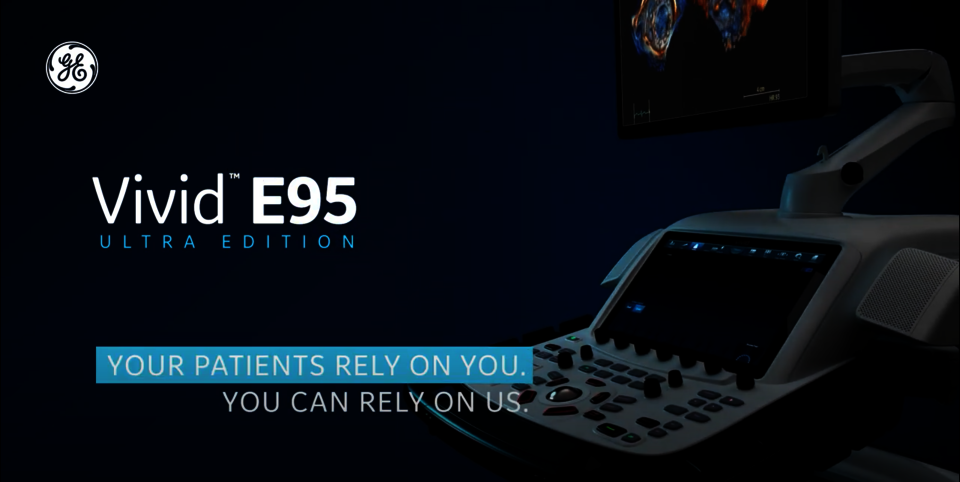
Vivid E95 Ultra Edition
Maximize your clinical confidence
Our ultrasound systems are designed to help you maximize your efficiency, achieve accurate measurements more easily and therefore minimize errors. Whether you are performing interventional or non-interventional examinations, or whether you are scanning adults or children: The Vivid E95 gives you a full suite of AI-based features to capture 4D ultrasound images quickly and efficiently.
Additionally, the combination of GE HealthCare's advanced probes and our powerful cSound software beamformer technology enables you to deliver ultrasound images of outstanding quality – for maximum clinical confidence.
Vivid E95 Ultra Edition
Powered by artificial intelligence. Elevated by you.
cSound ADAPT is key²
cSound ADAPT is based on our cSound software beamformer technology. It allows for continous beamforming optimization adapting to patient anatomy and probe position – up to 100 times per second. Image quality is thus improved significantly, especially for patients who are difficult to scan.
Benefit from excellent detail and contrast resolution in your non-interventional exams using cSound ADAPT in combination with our 4Vc-D probe. For your interventional exams, you can always rely on our 6VT-D TEE probe to achieve ultra-high volume rates and uncompromising image quality.

What our users say
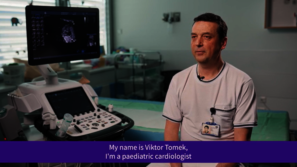
Usefulness of 3D TEE in the diagnosis of heart diseases in children
Product Highlights
Intelligent design for perfect ergonomics
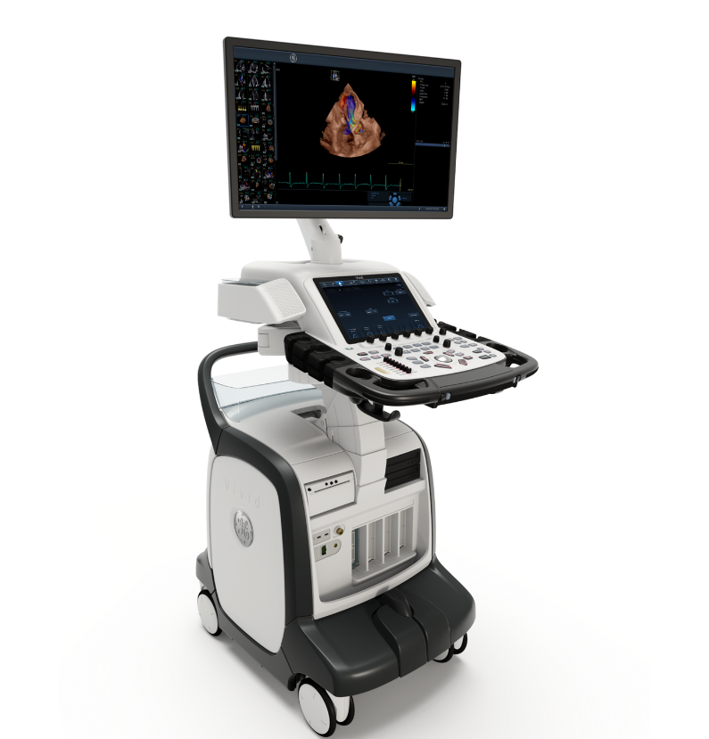
23.8" HDU-monitor
View your ultrasound images on our High Definition Ultrasound monitor (HDU) in ultra-high resolution and with excellent contrast.
Excellent operator interface
Vivid E95 comes with an electronically extendable, height- and side-adjustable control panel and is equipped with a 12" high-resolution LCD multi-touch screen.
Silent and economical
Silent fans and low heat dissipation guarantee reduced power consumption and minimum background noise.
Exceptional mobility
Thanks to its four swivel casters and its grab handles on the front and rear, you can easily move the Vivid E95 to wherever you need it. The electronic parking brake ensures maximum safety.
What our users say
Vivid E95 in prenatal fetal Echo diagnostics


References
- 3,5 times more processing power claim refers to the 2022 release of the Vivid portfolio. This Graphic Processing Unit is exclusively available for Vivid E95 and E90.
- cSound ADAPT: Continuous beamforming optimization, adapting to patient anatomy and probe position whitepaper - JB20851XX.
cSound Adapt is exclusively available for Vivid E95 and Vivid E90. - 9VT-D probe is exclusively available for Vivid E95 and Vivid E90 systems.The content herein refers to 2022 release of Vivid portfolio. Vivid Ultra Edition is released as of 25th August 2022.
* Ultra Edition is not a product name, it refers to the 2022 release of the Vivid portfolio.
Ultra fast. Ultra precise. Ultra efficient.
Shorter examination times. Increased precision.
Artificial intelligence algorithms enable the Vivid E95 Ultra Edition* to reduce manual steps and help improve the efficiency of your workflow through automated processes.
As our benchmark system for cardiovascular ultrasound, Vivid E95 is equipped with several imaging functions that are unique across the Vivid product family. Flexilight for example allows the photorealistic illumination of rendered 4D images, while 4D Auto TVQ enables fast, reproducible and accurate 4D visualization and quantification of the tricuspid valve.
Up to 80% fewer klicks¹ thanks to AI Auto Measure 2D
AI Auto Measure 2D semi-automatically measures left ventricle diameters from the parasternal long axis (PLAX). In this process, artificial intelligence algorithms recognize an end-diastolic and end-systolic image from an acquired parasternal long axis.
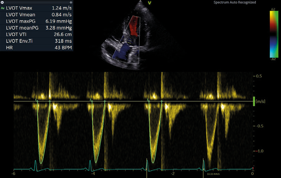
Cardiac Auto Doppler with AI Auto Measure Spectrum Recognition
With the power of AI, a wide range of Doppler measurements can be completed with two clicks: Freeze – Measure. A Doppler trace and full set of associated measurements will instantly appear on the screen. So you will need up to 93% fewer keystrokes.²
EASY AFI LV with View Recognition
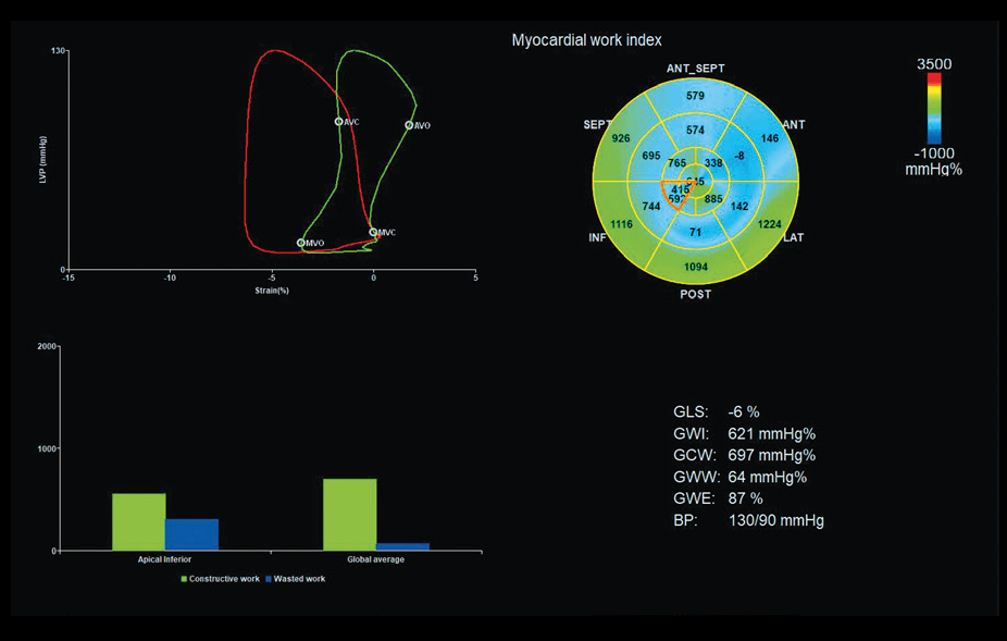
Easy AFI is the third generation of the 2D speckle tracking analysis tool for quantitative assessment of global and segmental myocardial function, which additionally detects the individual apical sectional planes fully automatically.
Through new AI-based detection algorithms, EASY AFI allows you to perform strain analysis without manual interaction. All you have to do is initiate the analysis and approve the results with one click – you will receive the results in only 15 seconds.³

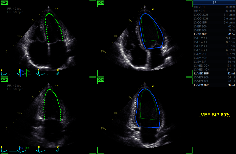
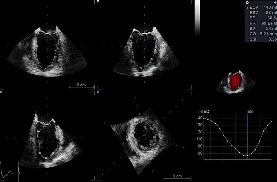
EASY AutoEF: Results with just one click⁴
In addition to our AutoEF quantification tool, which already automatically recognises the necessary sections (4-chamber and 2-chamber view) and selects them for the calculation, you can now determine the EF without manual interaction using our AI-based Auto-ROI recognition algorithms. All you have to do is initiate the analysis and approve the results with just one click.
Clinical Images
AFI – Automated Function Imaging


What our users say
Usefulness of speckle tracking AFI and Myocardial Work analysis in pediatrics
Clinical Images
Advanced 4D imaging capabilities


Clinical Images
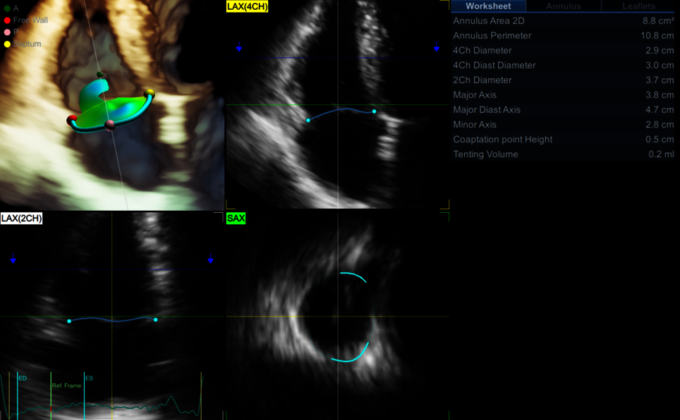
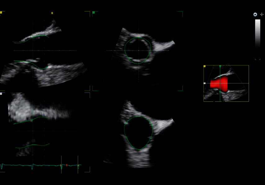
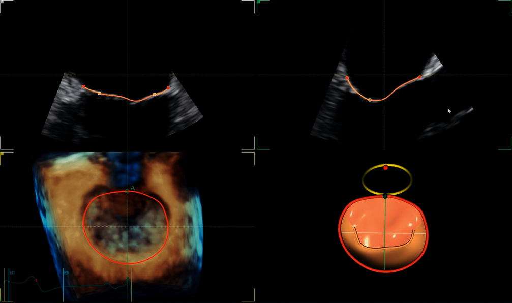
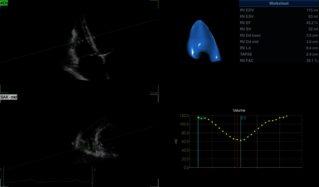
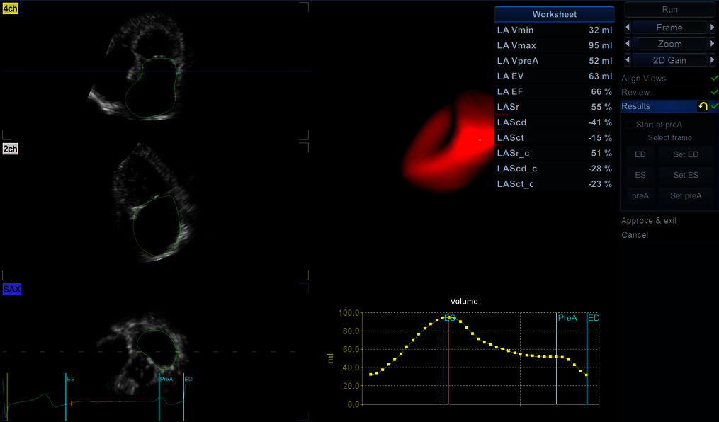
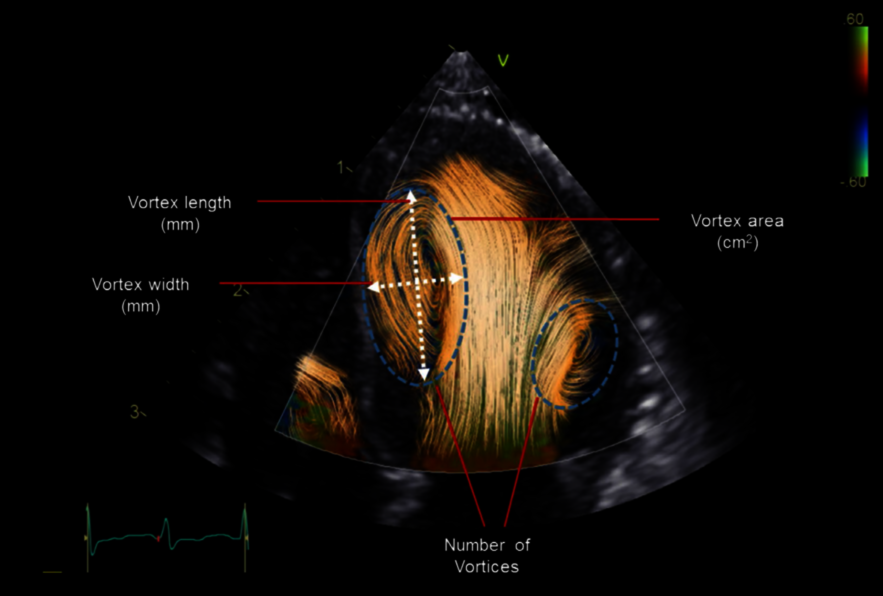
Ventricle and valve quantification in 4D


Clinical Images
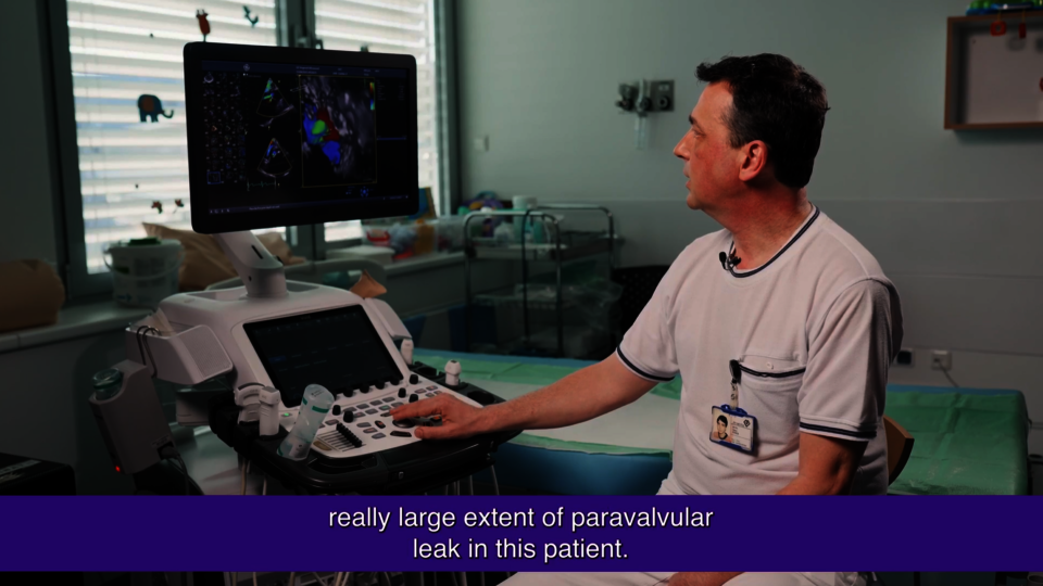
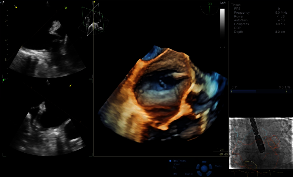
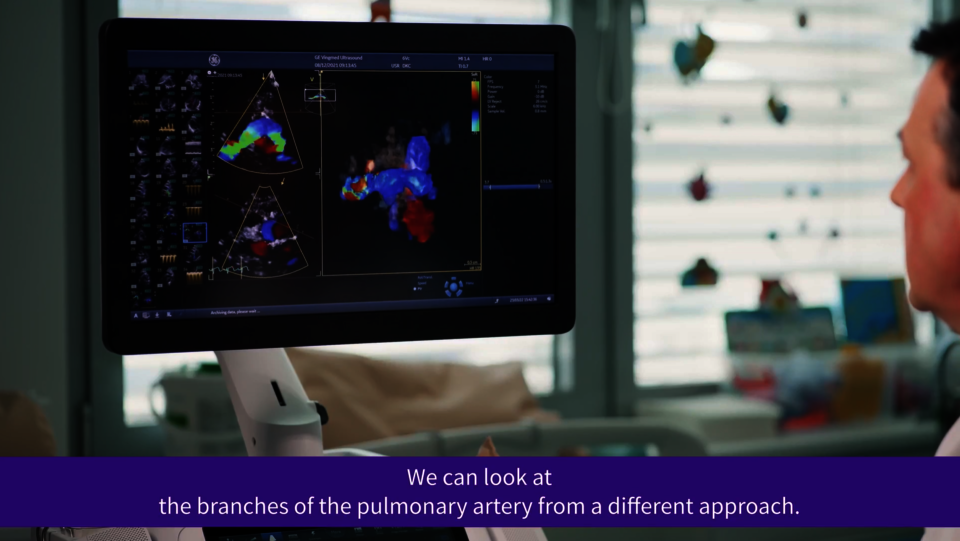
Interventional echocardiography


Clinical Expert’s perspective - Prof. Marcin Fijałkowski, MD, PhD
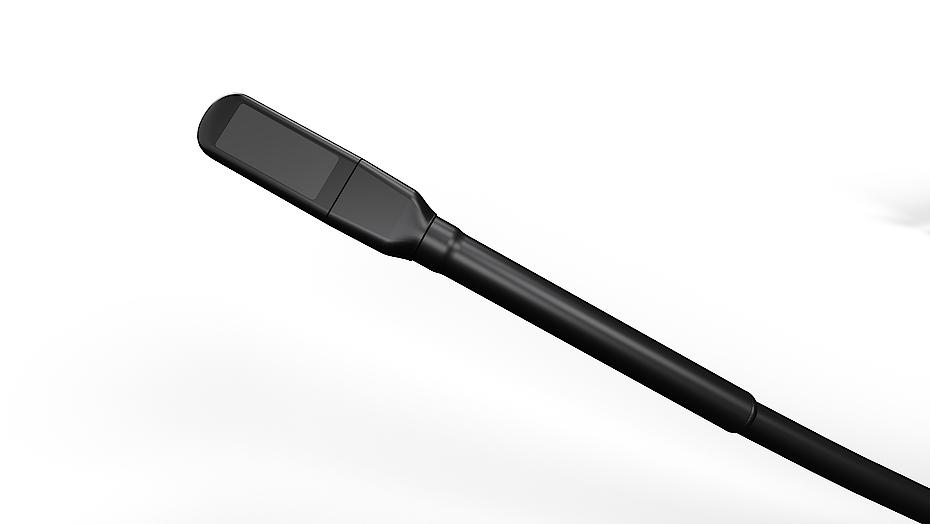
9VT-D mini 3D TEE Probe – Key Support in the Therapy of Challenging Patients

Clinical Images
Advanced probe technology for 2D and 4D pediatric ultrasound imaging
What our users say
Usefulness of 3D TEE in the diagnosis of congenital heart diseases in children
References
- The Role of AI in Streamlining Echocardiography Quantification White Paper, Kristin McLeod, Jurica Sprem JB20789XX.
- Based on results of time and motion study conducted by GE “JB49055XX - Cardiac Auto Doppler”; study results indicated time savings related productivity increase up to ~8 on an annual basis for a facility per sonographer.
- Time to strain measurement result may vary with heart rate, frame rate and Vivid system. Verification of performance done by GEHC clinical application specialists using Vivid system (DOC2739637)
- Easy AutoEF is restricted for use with adult TTE on GE Healthcare raw B-mode data loops of the LV. Easy AutoEF does not support left ventricles with septal bulge.
* Ultra Edition is not a product name, it refers to the 2022 release of the Vivid portfolio.
Dive into the world of Vivid E95 Ultra Edition*
In this section you will find valuable and detailed information on Vivid E95 and its features. Choose from brochures, whitepapers and case studies that offer you detailed insights into the functioning and application areas of our ultrasound system.


References
- 9VT-D probe is exclusively available for Vivid E95 and Vivid E90 systems. The content herein refers to 2022 release of Vivid portfolio. Vivid Ultra Edition is released as of 25th August 2022.
* Ultra Edition is not a product name, it refers to the 2022 release of the Vivid portfolio.