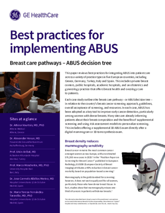
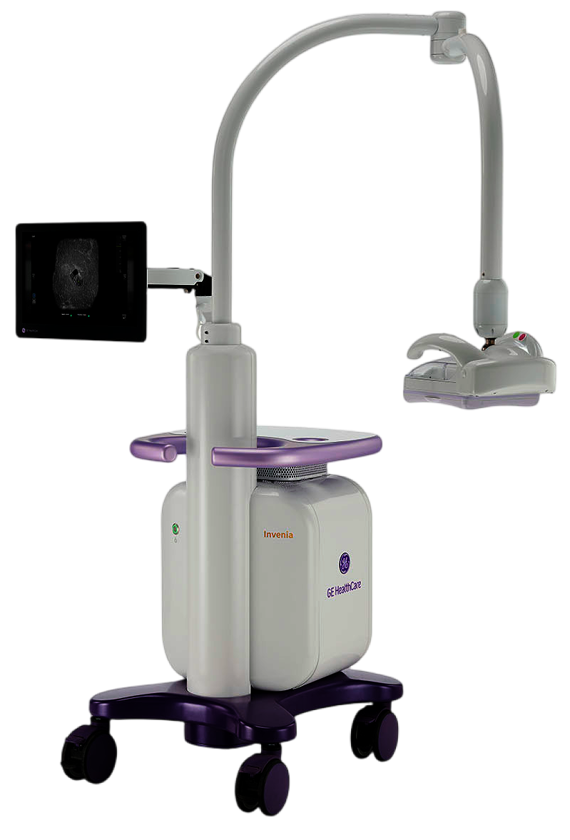
Invenia ABUS Premium
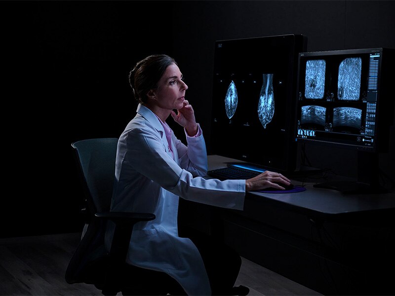
Invenia™ ABUS Premium provides the most innovative, patient-friendly, efficient and AI-driven 3D breast ultrasound designed to deliver optimal outcomes.
At a glance
Impactful screening solution
Improve breast cancer detection by 55% over mammography alone in dense breasts¹
Productivity powered by AI
Boost clinical confidence and efficiency with AI-driven scanning and reading
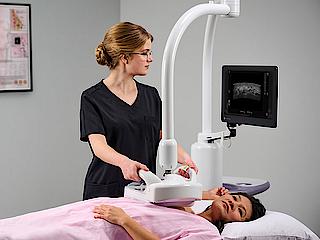
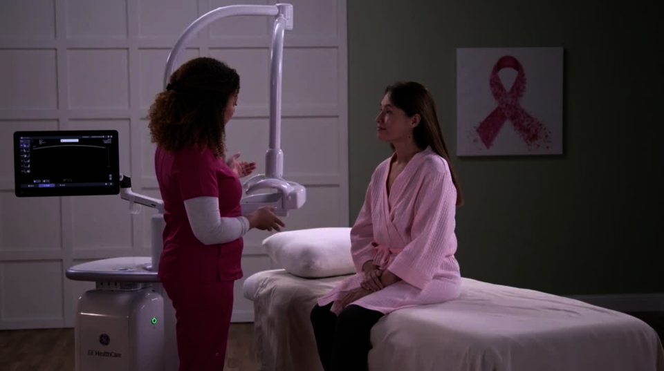
Outstanding patient comfort
Explore a non-invasive and patient-friendly breast ultrasound experience
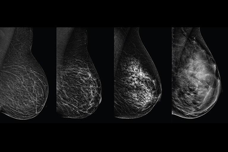
Why breast density matters
Dense breast tissue and cancer appear white on a mammogram, potentially camouflaging small cancers, and in fact, up to 50% of breast cancers may be missed in extremely dense breasts.2
Invenia ABUS Premium
Product Highlights
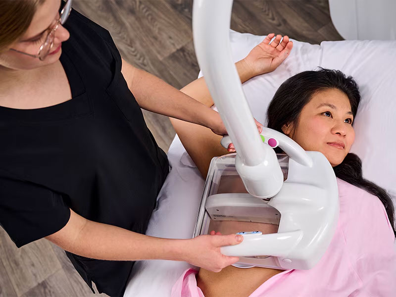
Reverse Curve™ transducer
15.3 cm Reverse Curve transducer follows the breast’s natural contour for outstanding patient comfort and full contact.
AI Assistant to improve clinical confidence
The Invenia ABUS Viewer with AI Assistant** integrates FDA-approved AI tools to enhance ABUS 3D dataset reviews, seamlessly incorporating intelligent algorithms to assist in characterizing breast lesions, providing a second opinion for added confidence.
Programmable hot keys
Programmable hot keys enable users to define commonly used functions to help reduce keystrokes.
Three-view layout option
Three-view layout option displays a synchronized view of multiple acquisitions on a single screen, allowing physicians to efficiently evaluate and cross reference areas of interest from multiple angles and increase diagnostic confidence.
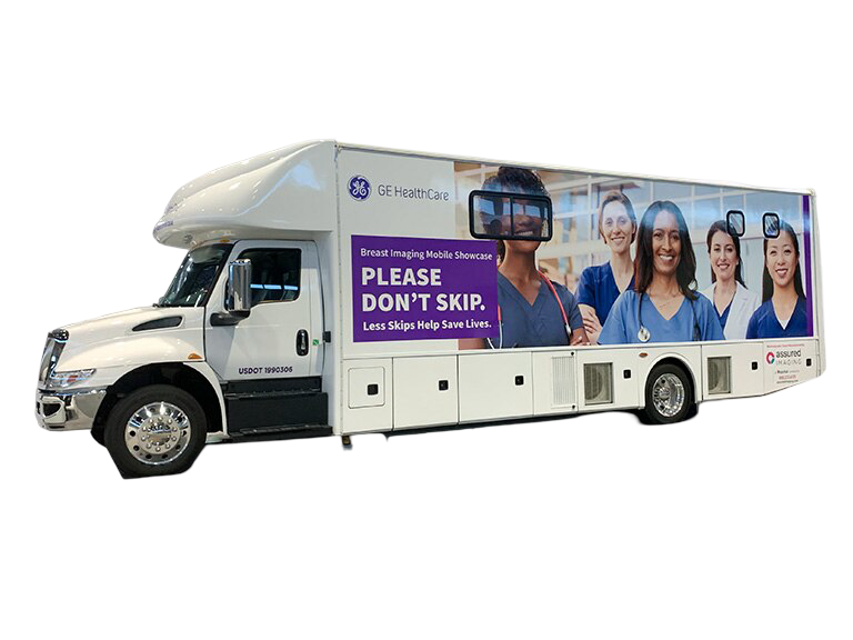
Bring the power of ABUS to your patients
The Mobile Invenia™ ABUS solutions let you bring extraordinary patient care to more places with the Mobile ABUS Fixation Bracket***. With 100% of women recommending ABUS to their best friend5, why not bring it directly to patients everywhere?
Invenia ABUS is an important pillar in the comprehensive Breast Care Pathway
Screening
Exact lesion localization and double reading capability empower ABUS in early detection
Diagnostic
Virtual rescans and multiplanar readings of the entire breast support case management
Planning
Offers a 3D view of the entire breast for precise and effective therapy planning
Monitoring
Compare reproducible ABUS volumes to priors to help monitor therapy response


Disclaimers
*Compared to Invenia ABUS 2.0.
**AI Assistant third-party AI tool powered by Koios DS Breast. AI Assistant enables seamless integration of GE HealthCare and third-party AI tools powered by Koios DS™ Breast.
***The Invenia ABUS device is not designed for use in a mobile environment unless it is installed using the Mobile ABUS fixation system.
References
- Brem RF, Tabár L, et.al. Radiology. 2015 Mar; 274(3): 663-73.
- Kolb et al, Radiology, Oct 2002;225(1):165-75.
- Pisano et al. NEJM 2005; 353: 1773.
- Engmann NJ, et al, JAMA Oncol. 2017;3(9):1228-1236.
- Shah et.al. Journal of Diagnostic Medical Sonography DOI: 10.1177/8756479313476920 2013.
- Barinov, et al. Impact of Data Presentation on Physician Performance Utilizing Artificial Intelligence-Based Computer- Aided Diagnosis and Decision Support Systems. J Digit Imaging (2018). doi.org/10.1007/s10278-018-0132-5.
- Wenhui Ren et. al, Elsevier Acad Radiol 2023; 30:S114
Features
The latest innovation in breast ultrasound
The Invenia ABUS Premium is designed for high patient throughput and extraordinary image quality to provide a great level of confidence. With its innovative design, it’s easy to use, reproducible, user-independent, standardized and allows reading anywhere.
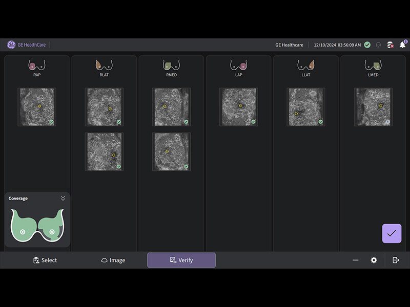
Verisound™ AI-powered solutions enhance confidence and speed workflow
Scan Quality Assessment provides an immediate, qualitative evaluation for proper breast coverage and positioning.
Auto Nipple Detection automatically offers nipple marker positioning to enable consistency and speed exams.
The novel Reverse Curve™ transducer is designed for enhanced performance
Its gentle 15.3 cm shape follows the breast's natural contour, enhancing patient comfort* and ensuring full contact for comprehensive coverage. The improved image quality* increases diagnostic confidence for reading ABUS exams.
Excellent image quality and 40% faster scan speed
cSound Imageformer, software-based processing, produces exquisite, reproducible images by leveraging automatic focus at every pixel.
Fast Scan increases scan speed by 40%*, fostering improved efficiency and patient throughput.
Centralized reading everywhere with the Invenia ABUS Viewer with AI Assistant
Invenia ABUS Viewer’s efficient workflow, with AI Assistant**, allows physicians to quickly review, interpret, and archive exams, even remotely.
Decrease benign biopsies by up to 55% with Koios DS™ Breast6
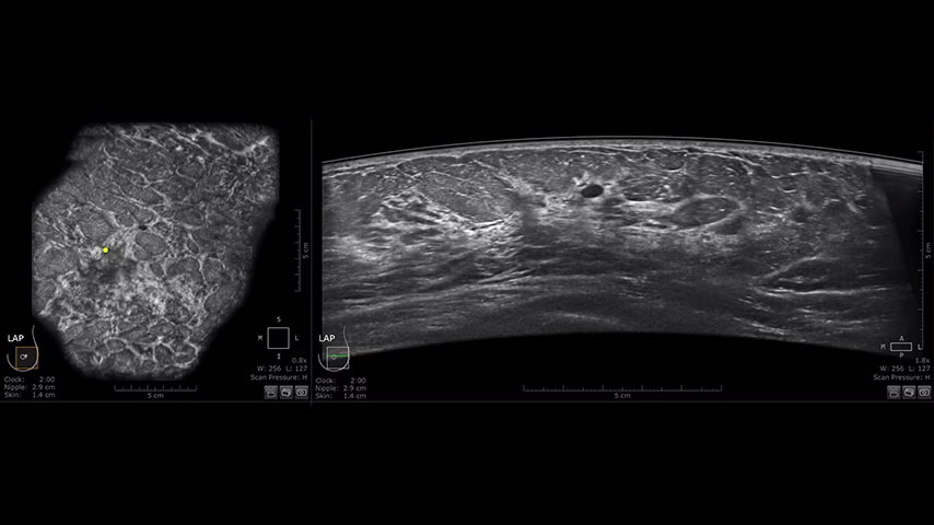
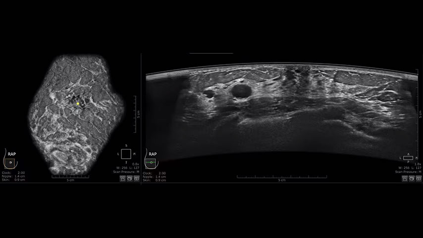
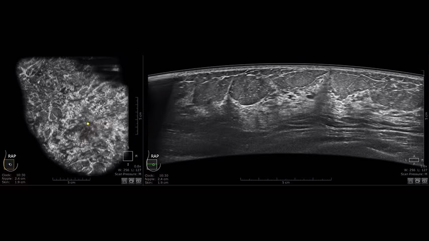
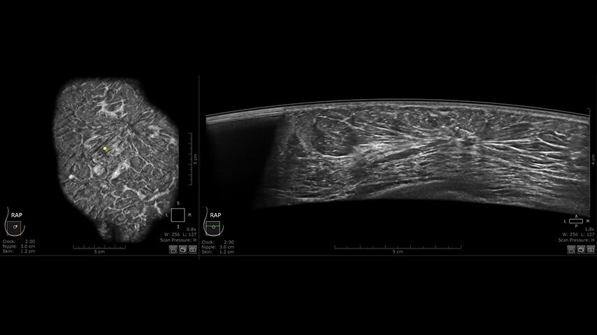
Enhanced image quality
The power of early detection with supplemental screening

References
- Brem RF, Tabár L, et.al. Radiology. 2015 Mar; 274(3): 663-73.
- Kolb et al, Radiology, Oct 2002;225(1):165-75.
- Pisano et al. NEJM 2005; 353: 1773.
- Engmann NJ, et al, JAMA Oncol. 2017;3(9):1228-1236.
- Shah et.al. Journal of Diagnostic Medical Sonography DOI: 10.1177/8756479313476920 2013.
- Barinov, et al. Impact of Data Presentation on Physician Performance Utilizing Artificial Intelligence-Based Computer- Aided Diagnosis and Decision Support Systems. J Digit Imaging (2018). doi.org/10.1007/s10278-018-0132-5.
- Wenhui Ren et. al, Elsevier Acad Radiol 2023; 30:S114