At a glance
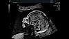
Effortless imaging
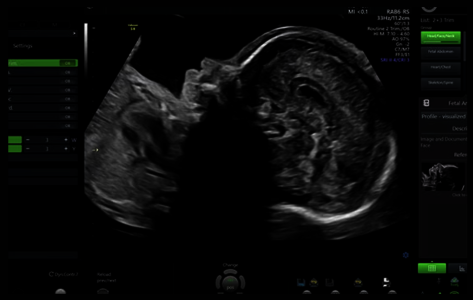
Capture images quickly and confidently with our Voluson Core Architecture. Just place the transducer and start scanning to acquire images with minimal manipulation.
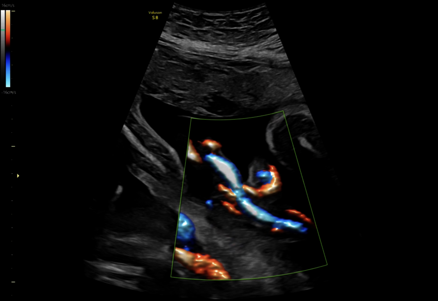
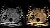
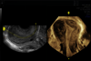
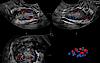
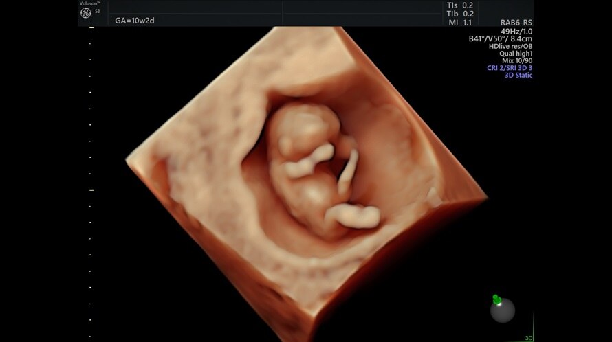
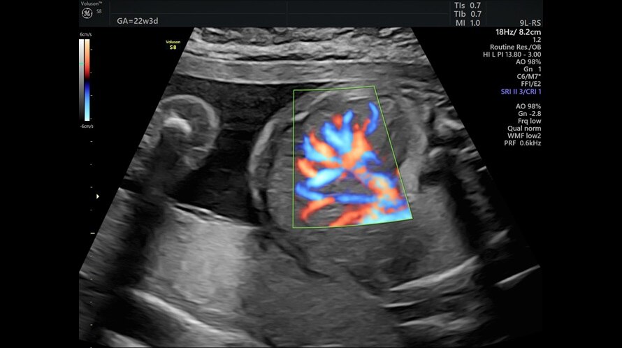
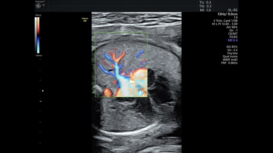
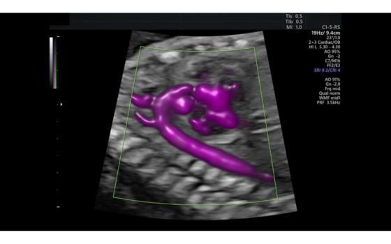
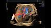
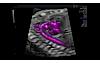
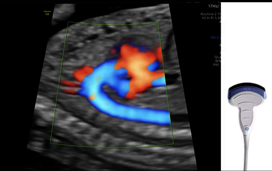
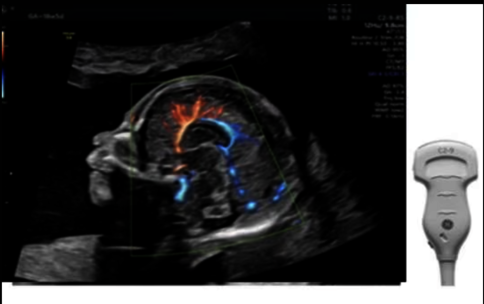
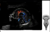
Exceptional color sensitivity
Enhance visualization of small vessels and borders of the fetal heart with Radiantflow™; powering greater color sensitivity for a more dynamic 3D-like appearance.
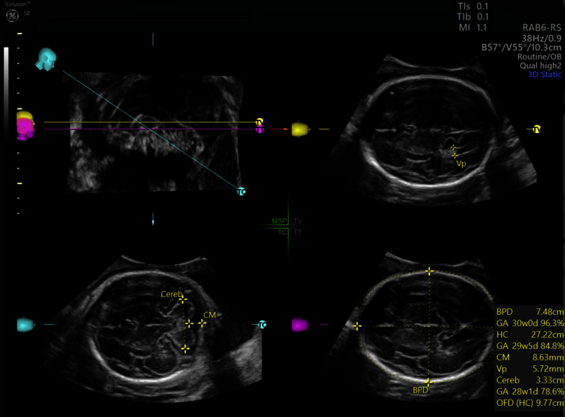
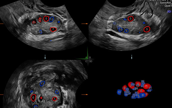
Workflow efficiency
Thanks to our advanced AI-based automation tools and the simple and intuitive operation of the ultrasound system, you can work with greater ease and efficiency.
More flexibility
With advanced ergonomics and features that focus on your dedicated clinical specialty, the Voluson S8 Touch offers both comfort and choice at an affordable price.
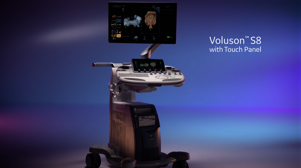
Voluson S8 Touch
Simple workflows. Excellent patient care.
Your busy practice demands a reliable ultrasound system that speeds you up. Boost your efficiency and productivity with the Voluson S8 Touch – the smart investment that allows you to set the pace.
Transform your workflow and save valuable time with fast, clear, and consistent imaging and automation tools from our premium segment. Experience more flexibility with an ultrasound system that fits your clinical specialty and your budget. Furthermore, our Voluson ecosystem provides you with comprehensive service and support as well as easy and efficient reporting and data management.
Voluson S8 Touch
Extraordinary efficiency: More time for you and your patients
Product Highlights
Pioneering in operational comfort and productivity
Designed for simplicity and comfort
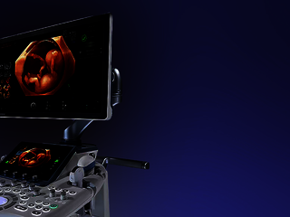
Voluson S8 Touch combines the ease-of-use of a tablet PC with the comfort of an articulating 23" widescreen monitor with adjustable layout formats.
Ergonomic operator interface
Voluson S8 Touch is equipped with a large 10.1" touch panel with Voluson xTouch functionality for an intuitive volume navigation experience. The panel's horizontal and vertical position can be adjusted with just one touch of a button.
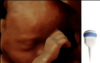
Advanced probe technology
Voluson S8 Touch comes with an ultra-light and high-frequency abdominal probe that is ideal for difficult examination conditions. In addition, a special transducer with matrix array technology is available for advanced mamma diagnostics.
Fast and secure data management
Use the state-of-the-art communication technologies of Voluson S8 Touch to quickly exchange data with patients and colleagues and to archive your images easily.
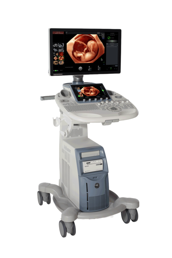
Mobile and flexible
Equipped with four casters, the lightweight ultrasound system moves easily to wherever you need it. The integrated battery pack allows for up to 20 minutes of scanning.


Expert ultrasound for your practice
Expert imaging capabilities made affordable
From routine ultrasound examinations to advanced, detailed diagnostics at the highest level: The Voluson S8 Touch was designed to obtain critical answers fast. Built on Voluson Core Architecture, the ultrasound system incorporates a legacy of innovation to deliver excellent imaging. At the same time, effort for analysis and reporting is reduced thanks to various built-in automation tools and sophisticated software applications. Choose from a wide range of functions to perfectly adapt the system to your clinical specialties.
Voluson technology
Discover Voluson technology for advanced ultrasound imaging
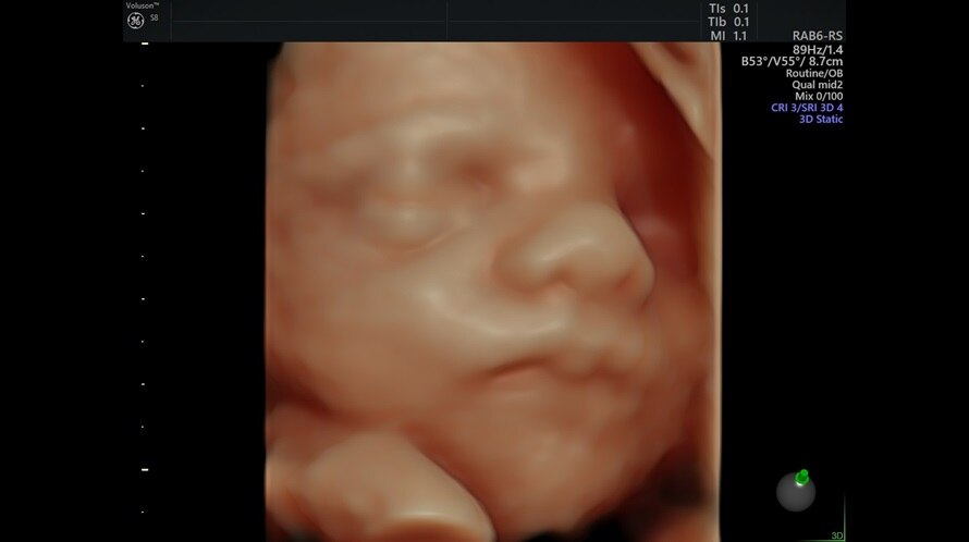


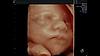
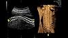
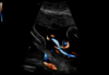
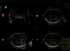
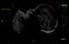
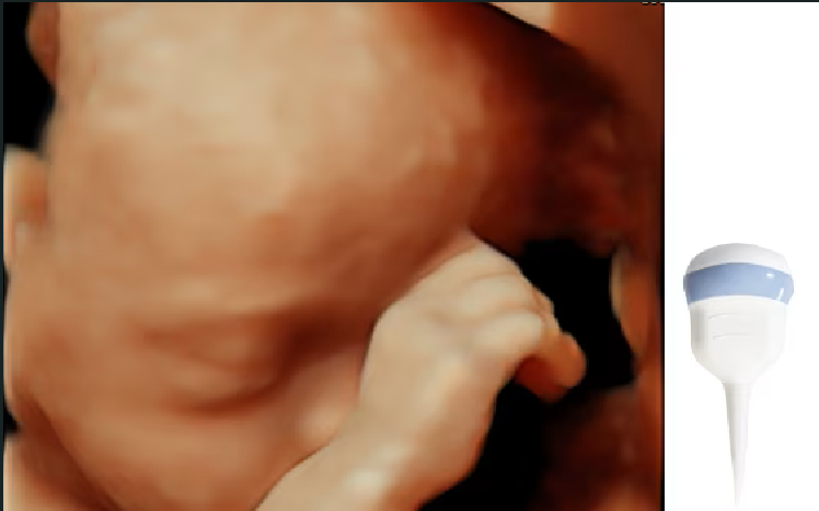
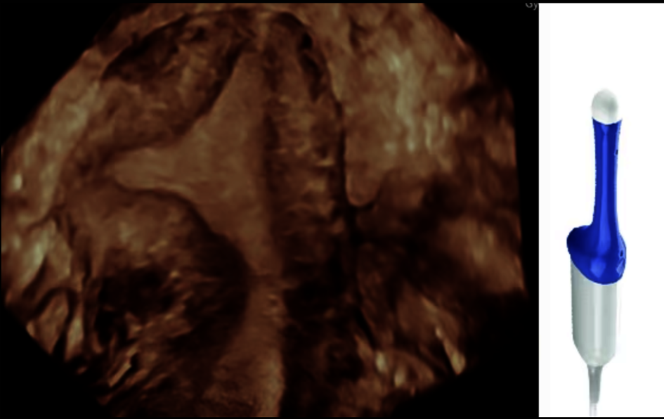
Clinical images
Voluson S8 Touch at work
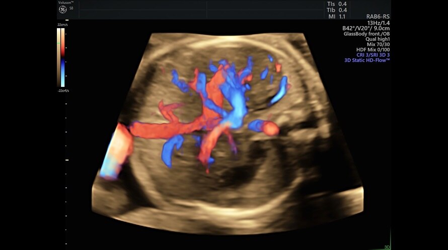
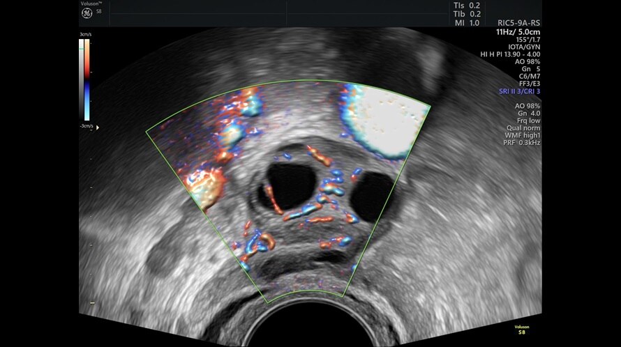
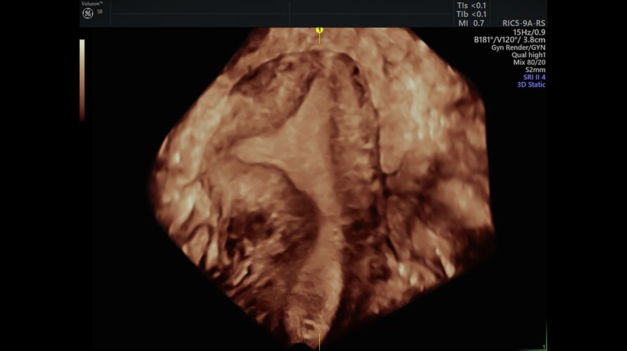
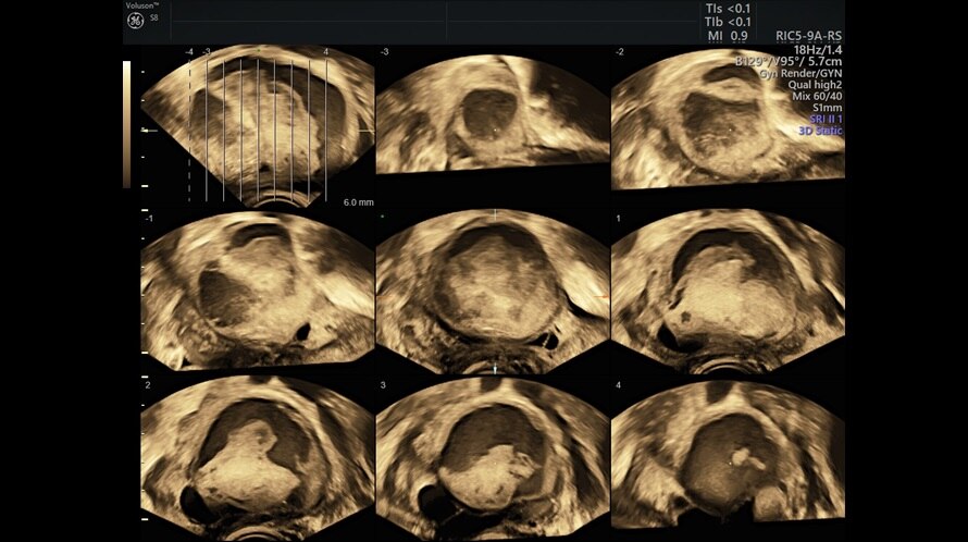
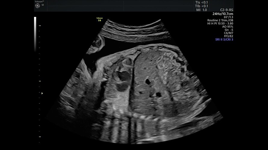
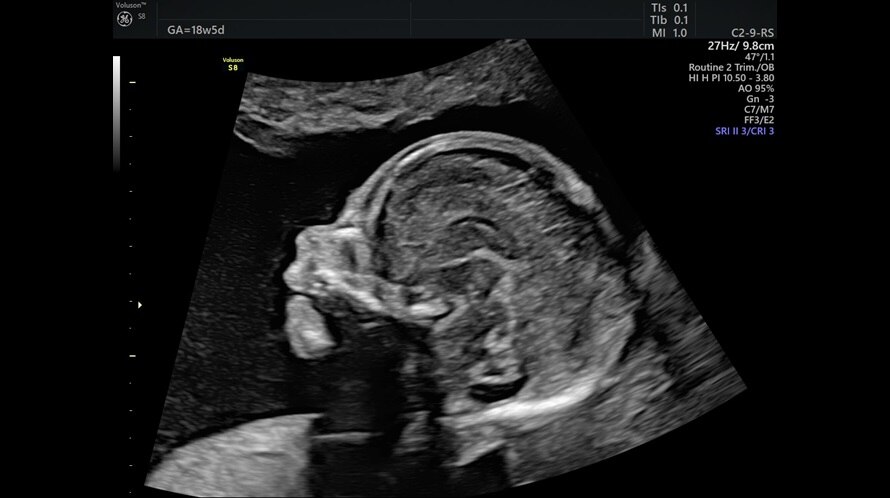
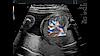


Clinical Images
Voluson probe technology for extraordinary ultrasound images


Dive into the world of Voluson S8 Touch
In this section you will find valuable and detailed information on Voluson S8 Touch and its features. Choose from brochures, whitepapers and case studies that offer you detailed insights into the functioning and application areas of our ultrasound system.