Voluson™ SWIFT
Welcome to a remarkably different ultrasound experience.
Transform the way you work – with Voluson SWIFT.
At a glance
This is simplicity
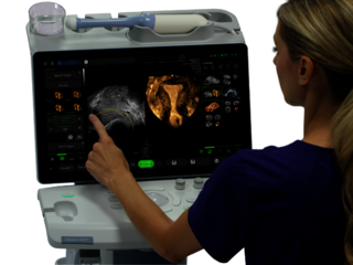
With a large touchscreen and a select group of hard keys, Voluson SWIFT incorporates the same functionality as the devices we use every day.
This is adaptability
Designed to help remove some of the obstacles that consistently slow you down. Streamline your workflow by leveraging the power of AI and automation.
This is efficiency
Highly customizable, and designed to simplify every interaction. Make it yours by personalizing the menus and workflows, and by customizing presets.
This is defining value
Expect more from your system. More services, support and partnership. Voluson SWIFT is a technologically advanced platform built for the future.
Voluson SWIFT
This changes everything
Uniquely intuitive and customizable, we’ve reimagined the user experience – for a new level of ease and efficiency. Radically different in look and feel, Voluson SWIFT is designed to simplify every interaction. Feel like an experienced user from the beginning with artificial intelligence and automated tools that expand your capabilities and inspire confidence.
Voluson SWIFT
Discover the future of women's health
Product Highlights
Increase precision and efficiency of your ultrasound exams – your way
Perfectly adaptable to your needs
Voluson SWIFT perfectly adapts to your workflow. You can personalize the menus, customize measurements and annotations and set up individualized user presets. All with just a few clicks.
Equipped with artificial intelligence
By leveraging the power of artificial intelligence, Voluson SWIFT allows you to save valuable time on acquisition, analysis, and reporting. Easy-to-use automation tools can streamline your workflow and increase consistency.
Easy reporting and data management
Use the Tricefy™ software of your Voluson SWIFT to archive your ultrasound images easily and to share them instantly with patients and colleagues.
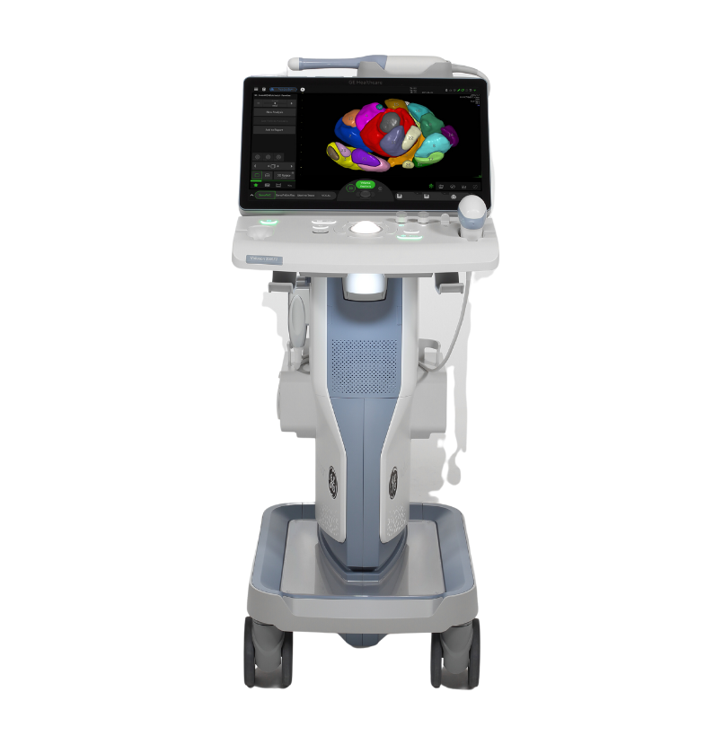
Perfect ergonomics
With its optimised key layout and simple touchscreen navigation, Voluson SWIFT allows for maximum efficient handling. The number of hard keys and solid surfaces has been reduced to a minimum, making cleaning and disinfection a matter of moments.
Always ready to scan
The built-in rechargeable battery ensures uninterrupted imaging wherever you are.
Experience support excellence
Leverage GE HealthCare's entire ecosystem of support for peace of mind – now and in the future.
- Count on rapid, responsive service to keep your ultrasound system up and running at the highest level.
- Access remote support from our technical and clinical experts to help identify and resolve issues in real-time.
- Use expert tools to manage the health of your ultrasound system and transducers.
- Take advantage of advanced data analytics to draw out key insights.


The future of women's health
Imaging technology that changes everything
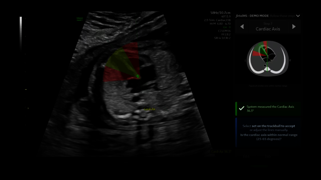
At Voluson, we are constantly innovating to improve image quality and create specialized clinical tools to help you see more anatomical detail with greater confidence, enabling you to provide the best possible patient care.
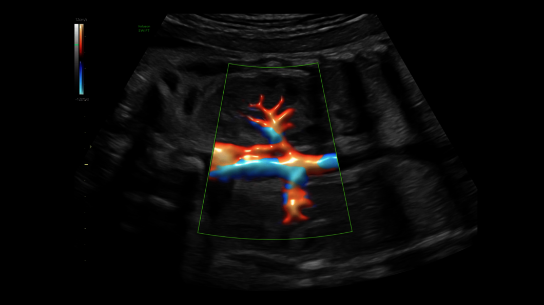
That's exactly why Voluson SWIFT is equipped with a comprehensive set of AI tools and Sono-Automation technologies that both reduce complexity, while increasing efficiency and consistency of your exams. HDlive™ technologies allow for ultrasound imaging with unprecedented clarity, while Radiantflow™ takes the color Doppler to a whole new level.
Voluson technology
Discover Voluson technology for advanced ultrasound imaging


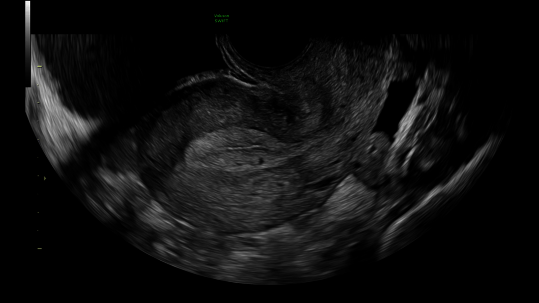
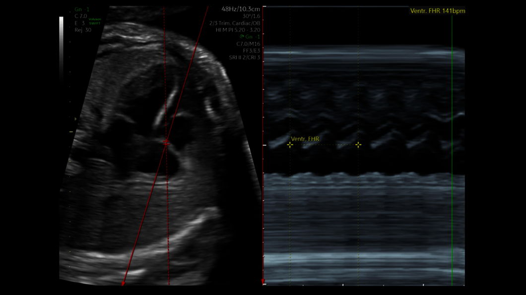
Clinical images
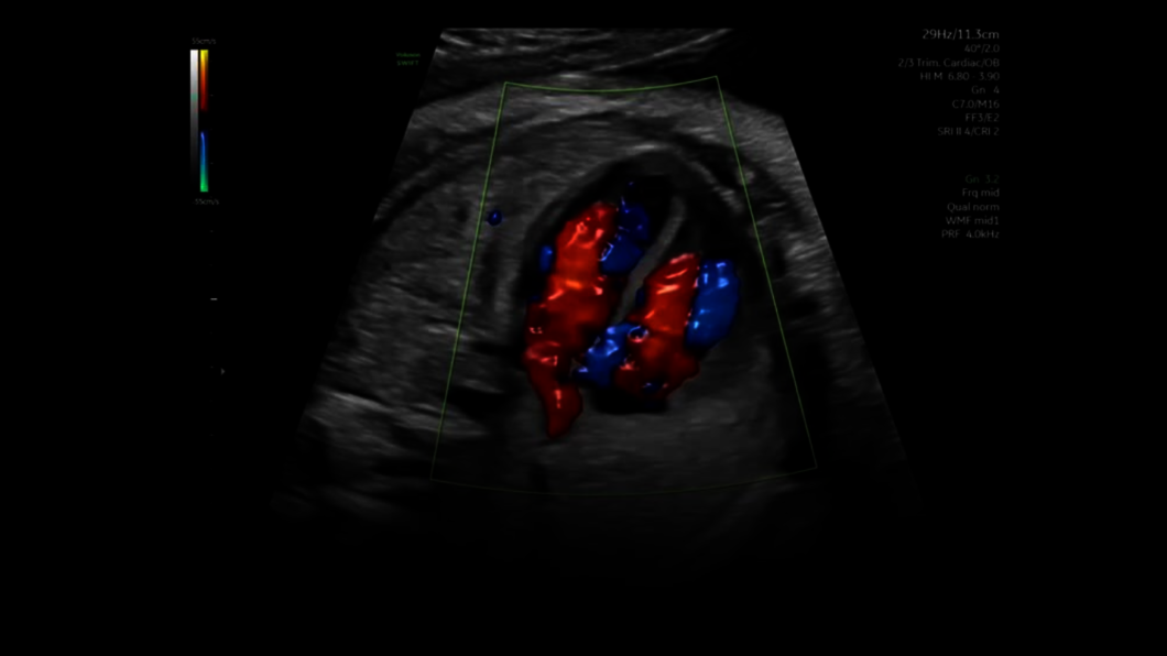
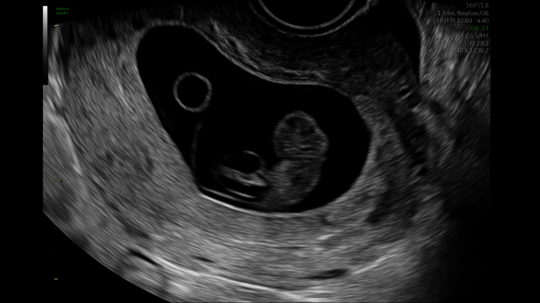
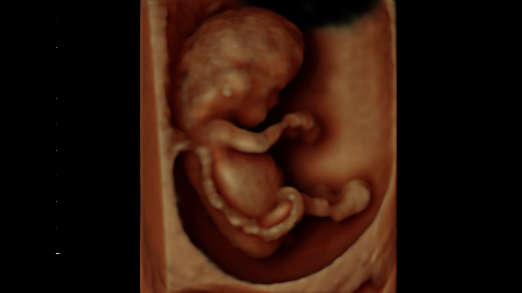
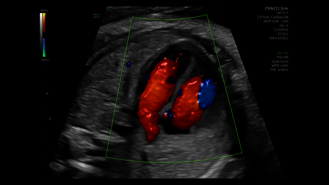
Obstetrics
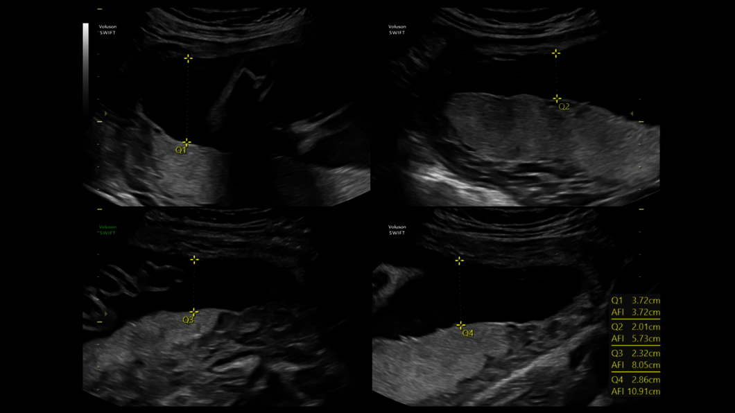
From early pregnancy to birth, the health of an expectant mother and her fetus is your priority. That’s why we make it our priority to ensure extraordinary imaging at every stage of pregnancy to confidently interrogate fetal anatomy and detect abnormalities earlier than ever before.


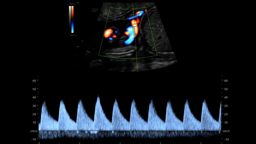
Clinical images
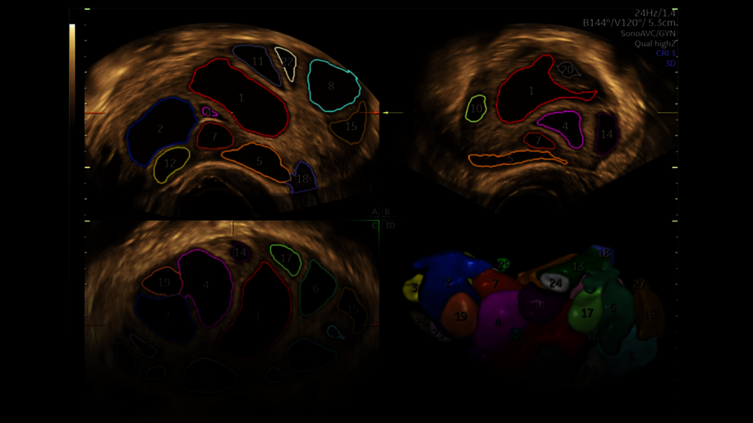
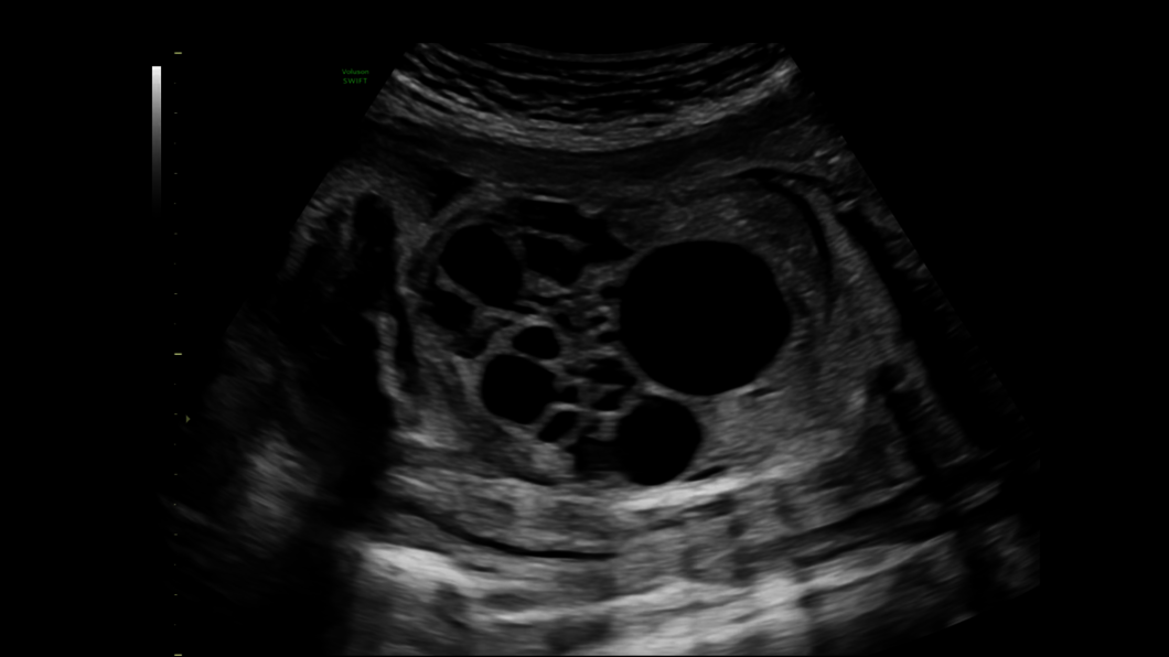
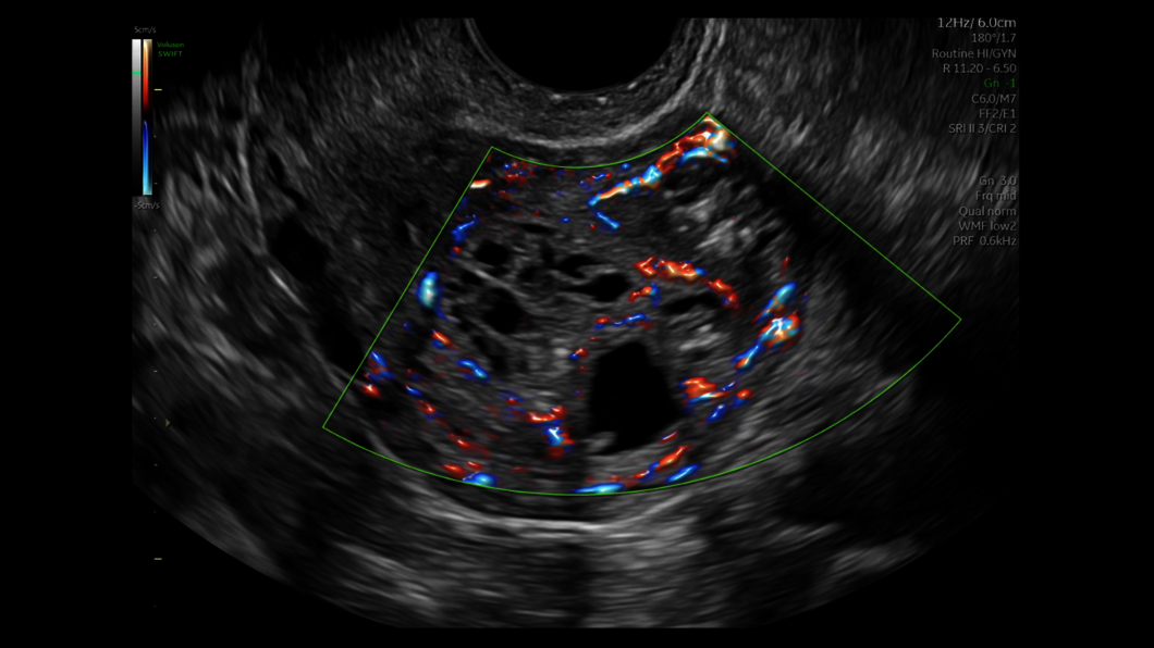
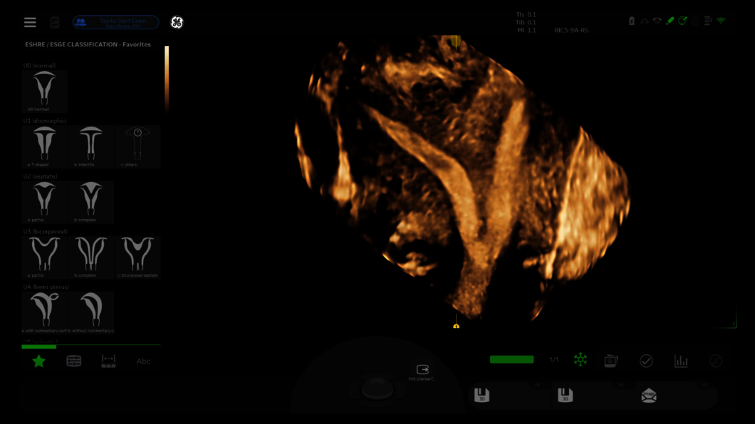
Gynecology
Ultrasound is often the first line of defense in diagnosing gynecological conditions. Pelvic pain, post-menopausal bleeding, genitourinary dysfunction and infertility can be confusing and concerning for your patients. They need answers and rely on your expertise for accurate diagnosis and management.


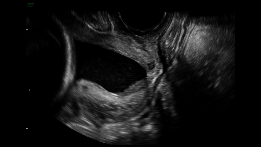
Clinical Images
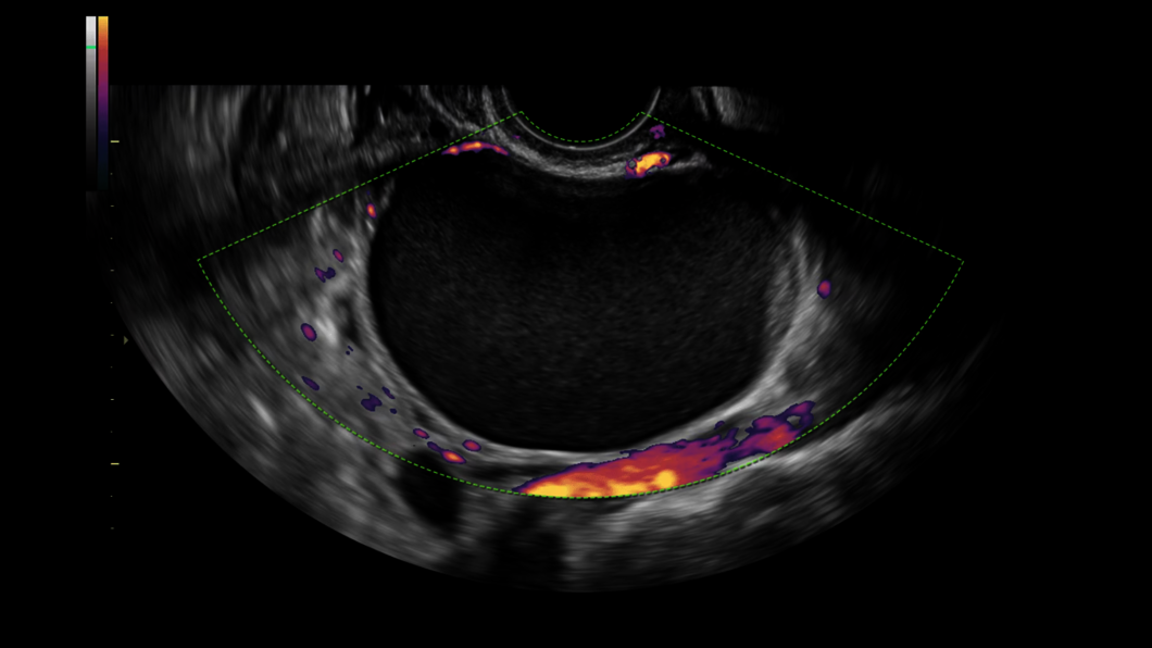
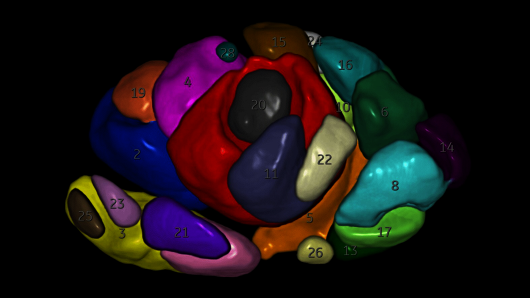
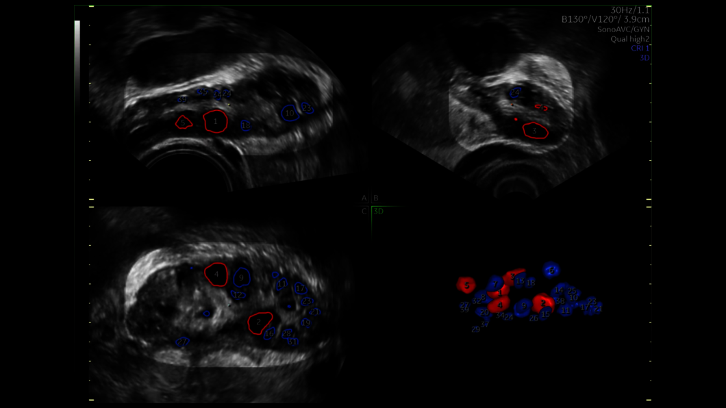
Assisted Reproductive Medicine
We understand the impact infertility has on your patients’ lives. That’s why our Voluson ultrasound systems continually push the boundaries of imaging to give you simple, yet innovative tools to help your patients achieve their dream of a successful pregnancy.


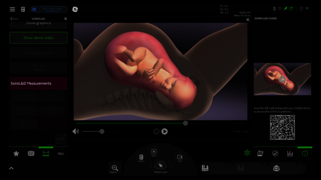
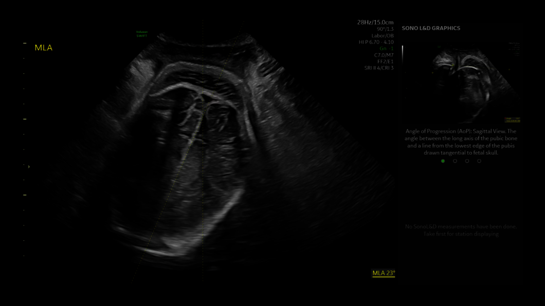
Clinical Images
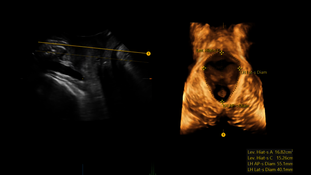
Labor & Delivery
The birthing process is filled with excitement, anticipation and sometimes uncertainty. With extraordinary imaging and innovative labor progression tracking, Voluson SWIFT can help you monitor labor progression as well as baby and mother wellness, helping you make more informed clinical decisions.


References
- HyCoSy / HyFoSy is not available in all countries.
Dive into the world of Voluson SWIFT
In this section you will find valuable and detailed information on Voluson SWIFT and its features. Choose from brochures, whitepapers and case studies that offer you detailed insights into the functioning and application areas of our ultrasound system.


